Deposition Date
2019-01-18
Release Date
2019-01-30
Last Version Date
2024-10-16
Entry Detail
PDB ID:
6NPZ
Keywords:
Title:
Crystal structure of Akt1 (aa 123-480) kinase with a bisubstrate
Biological Source:
Source Organism(s):
Homo sapiens (Taxon ID: 9606)
Expression System(s):
Method Details:
Experimental Method:
Resolution:
2.12 Å
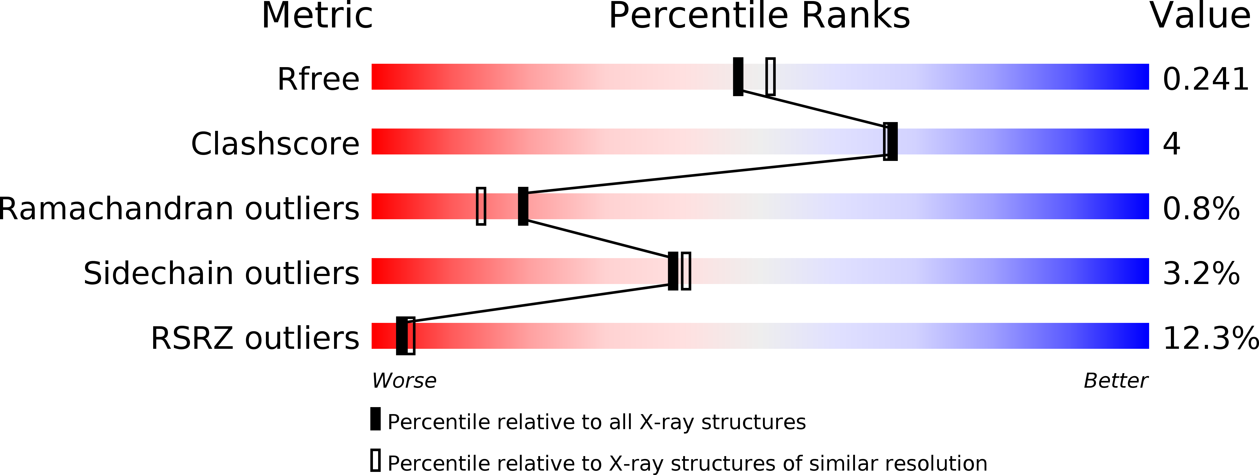
R-Value Free:
0.24
R-Value Work:
0.18
R-Value Observed:
0.18
Space Group:
P 1 21 1