6NMJ image
Deposition Date 2019-01-11
Release Date 2019-06-26
Last Version Date 2023-10-11
Entry Detail
PDB ID:
6NMJ
Title:
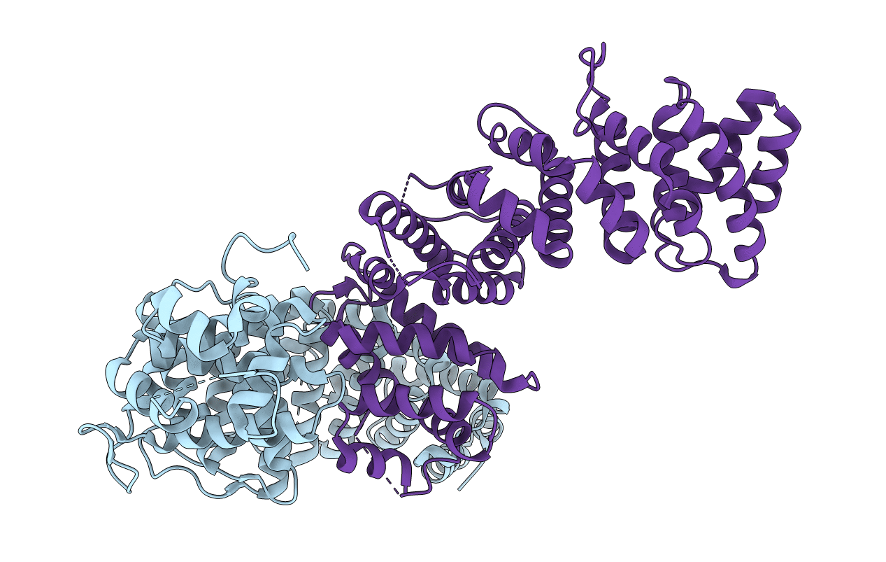
Crystal Structure of Rat Ric-8A G alpha binding domain, "Paratone-N Immersed"
Biological Source:
Source Organism:
Host Organism:
Method Details:
Experimental Method:
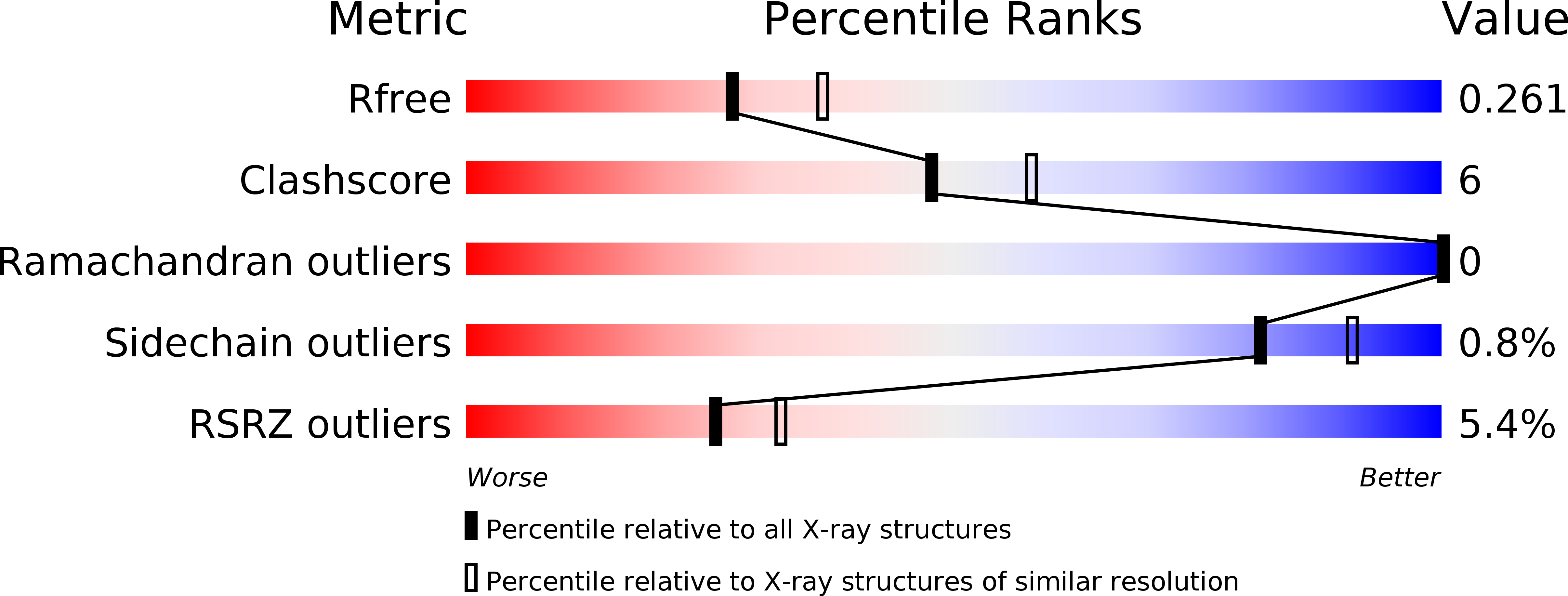
Resolution:
2.30 Å
R-Value Free:
0.25
R-Value Work:
0.20
R-Value Observed:
0.20
Space Group:
P 21 21 21
Macromolecular Entities
Polymer Type:polypeptide(L)
Molecule:Resistance to inhibitors of cholinesterase 8 homolog A (C. elegans)
Mutations:F232Y
Chain IDs:A, B
Chain Length:453
Number of Molecules:2
Biological Source:Rattus norvegicus
Primary Citation
Structure, Function, and Dynamics of the G alpha Binding Domain of Ric-8A.
Structure 27 1137 1147.e5 (2019)
PMID: 31155309 DOI: 10.1016/j.str.2019.04.013

Abstact

Ric-8A is a 530-amino acid cytoplasmic molecular chaperone and guanine nucleotide exchange factor (GEF) for i, q, and 12/13 classes of heterortrimeric G protein alpha subunits (Gα). We report the 2.2-Å crystal structure of the Ric-8A Gα-binding domain with GEF activity, residues 1-452, and is phosphorylated at Ser435 and Thr440. Residues 1-429 adopt a superhelical fold comprised of Armadillo (ARM) and HEAT repeats, and the C terminus is disordered. One of the phosphorylated residues potentially binds to a basic cluster in an ARM motif. Amino acid sequence conservation and published hydrogen-deuterium exchange data indicate repeats 3 through 6 to be a putative Gα-binding surface. Normal mode modeling of small-angle X-ray scattering data indicates that phosphorylation induces relative rotation between repeats 1-4, 5-6, and 7-9. 2D 1H-15N-TROSY spectra of [2H,15N]-labeled Gαi1 in the presence of R452 reveals chemical shift perturbations of the C terminus and Gαi1 residues involved in nucleotide binding.

Legend

Protein

Chemical

Disease

Primary Citation of related structures