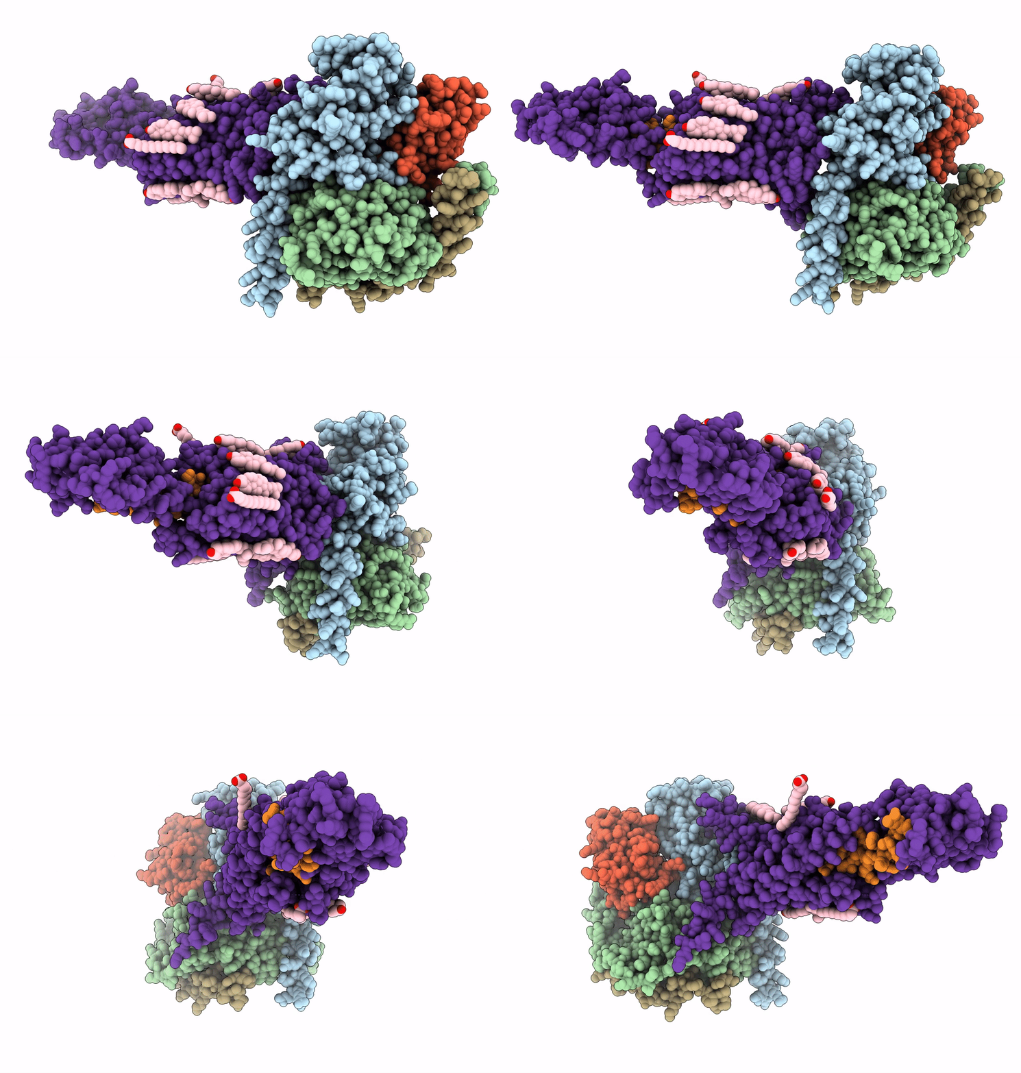
6NBH image
Entry Detail
PDB ID:
6NBH
EMDB ID:
Title:
Cryo-EM structure of parathyroid hormone receptor type 1 in complex with a long-acting parathyroid hormone analog and G protein
Biological Source:
PDB Version:
Deposition Date:
2018-12-07
Release Date:
2019-04-17
Method Details:
Experimental Method:
Resolution:
3.50 Å
Aggregation State:
PARTICLE
Reconstruction Method:
SINGLE PARTICLE
Macromolecular Entities
Polymer Type:polypeptide(L)
Description:Gs protein alpha subunit
Chain IDs:C (auth: A)
Chain Length:378
Number of Molecules:1
Biological Source:Bos taurus
Polymer Type:polypeptide(L)
Description:Guanine nucleotide-binding protein G(I)/G(S)/G(T) subunit beta-1
Chain IDs:D (auth: B)
Chain Length:345
Number of Molecules:1
Biological Source:Rattus norvegicus
Polymer Type:polypeptide(L)
Description:Guanine nucleotide-binding protein G(I)/G(S)/G(O) subunit gamma-2
Chain IDs:E (auth: G)
Chain Length:71
Number of Molecules:1
Biological Source:Bos taurus
Polymer Type:polypeptide(L)
Description:Nanobody-35
Chain IDs:F (auth: N)
Chain Length:126
Number of Molecules:1
Biological Source:synthetic construct
Polymer Type:polypeptide(L)
Description:Long-acting parathyroid hormone analog
Chain IDs:B (auth: P)
Chain Length:36
Number of Molecules:1
Biological Source:Homo sapiens
Polymer Type:polypeptide(L)
Description:Parathyroid hormone/parathyroid hormone-related peptide receptor
Mutations:G188A
Chain IDs:A (auth: R)
Chain Length:478
Number of Molecules:1
Biological Source:Homo sapiens
Primary Citation

Abstact

The parathyroid hormone receptor-1 (PTH1R) is a class B G protein-coupled receptor central to calcium homeostasis and a therapeutic target for osteoporosis and hypoparathyroidism. Here we report the cryo-electron microscopy structure of human PTH1R bound to a long-acting PTH analog and the stimulatory G protein. The bound peptide adopts an extended helix with its amino terminus inserted deeply into the receptor transmembrane domain (TMD), which leads to partial unwinding of the carboxyl terminus of transmembrane helix 6 and induces a sharp kink at the middle of this helix to allow the receptor to couple with G protein. In contrast to a single TMD structure state, the extracellular domain adopts multiple conformations. These results provide insights into the structural basis and dynamics of PTH binding and receptor activation.

Legend

Protein

Chemical

Disease

Primary Citation of related structures