6N9W image
Entry Detail
PDB ID:
6N9W
EMDB ID:
Title:
Structure of bacteriophage T7 lagging-strand DNA polymerase (D5A/E7A) and gp4 (helicase/primase) bound to DNA including RNA/DNA hybrid, and an incoming dTTP (LagS2)
Biological Source:
Source Organism:
Host Organism:
PDB Version:
Deposition Date:
2018-12-04
Release Date:
2019-03-06
Method Details:
Experimental Method:
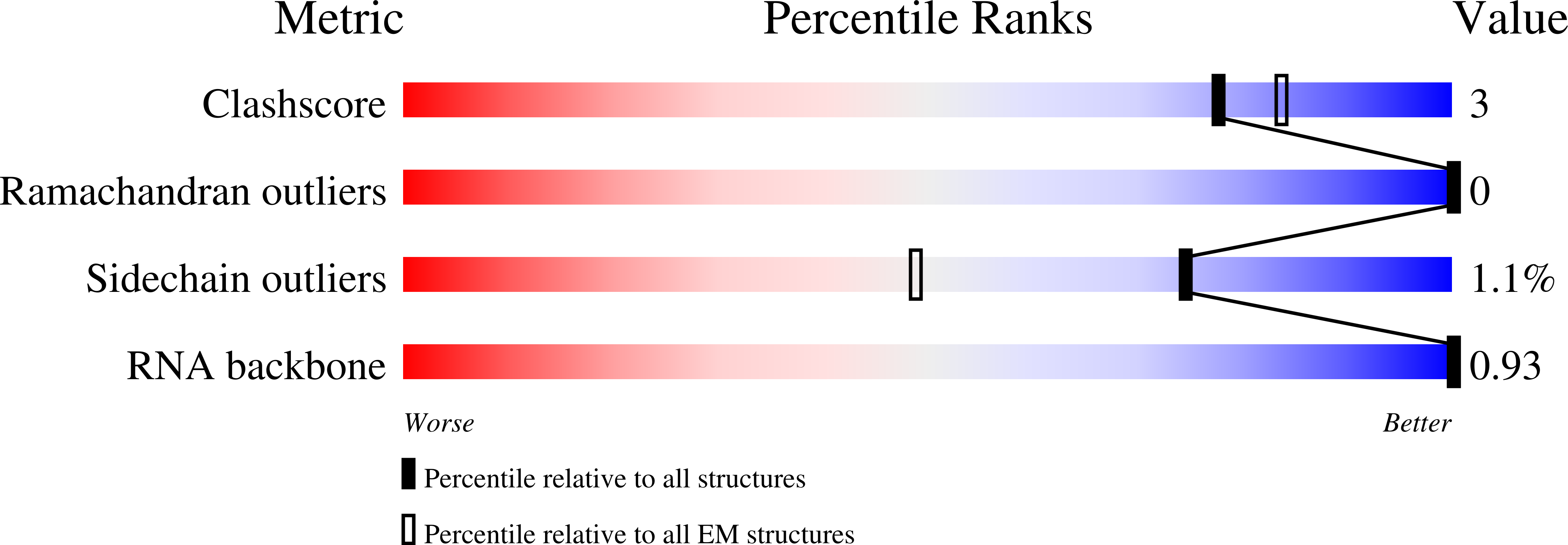
Resolution:
4.00 Å
Aggregation State:
PARTICLE
Reconstruction Method:
SINGLE PARTICLE
Macromolecular Entities
Polymer Type:polypeptide(L)
Description:DNA primase/helicase
Mutations:E343Q
Chain IDs:A, B, C, D, E, F
Chain Length:566
Number of Molecules:6
Biological Source:Enterobacteria phage T7
Polymer Type:polypeptide(L)
Description:DNA-directed DNA polymerase
Mutations:D5A, E7A
Chain IDs:G (auth: H)
Chain Length:704
Number of Molecules:1
Biological Source:Enterobacteria phage T7
Polymer Type:polyribonucleotide
Description:Primer
Chain IDs:H (auth: P)
Chain Length:6
Number of Molecules:1
Biological Source:Enterobacteria phage T7
Polymer Type:polydeoxyribonucleotide
Description:Template
Chain IDs:I (auth: T)
Chain Length:44
Number of Molecules:1
Biological Source:Enterobacteria phage T7
Primary Citation
Structures and operating principles of the replisome.
Science 363 ? ? (2019)
PMID: 30679383 DOI: 10.1126/science.aav7003

Abstact

Visualization in atomic detail of the replisome that performs concerted leading- and lagging-DNA strand synthesis at a replication fork has not been reported. Using bacteriophage T7 as a model system, we determined cryo-electron microscopy structures up to 3.2-angstroms resolution of helicase translocating along DNA and of helicase-polymerase-primase complexes engaging in synthesis of both DNA strands. Each domain of the spiral-shaped hexameric helicase translocates sequentially hand-over-hand along a single-stranded DNA coil, akin to the way AAA+ ATPases (adenosine triphosphatases) unfold peptides. Two lagging-strand polymerases are attached to the primase, ready for Okazaki fragment synthesis in tandem. A β hairpin from the leading-strand polymerase separates two parental DNA strands into a T-shaped fork, thus enabling the closely coupled helicase to advance perpendicular to the downstream DNA duplex. These structures reveal the molecular organization and operating principles of a replisome.

Legend

Protein

Chemical

Disease

Primary Citation of related structures