Deposition Date
2018-11-08
Release Date
2019-05-22
Last Version Date
2023-10-11
Entry Detail
PDB ID:
6N1K
Keywords:
Title:
Full-length human phenylalanine hydroxylase (PAH) in the resting state
Biological Source:
Source Organism(s):
Homo sapiens (Taxon ID: 9606)
Expression System(s):
Method Details:
Experimental Method:
Resolution:
3.06 Å
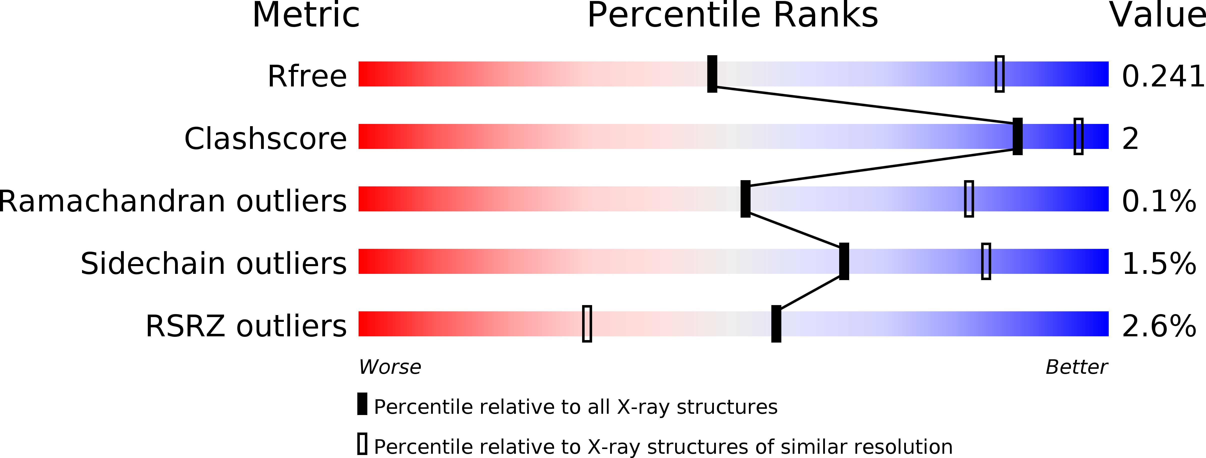
R-Value Free:
0.23
R-Value Work:
0.20
R-Value Observed:
0.20
Space Group:
P 1 21 1