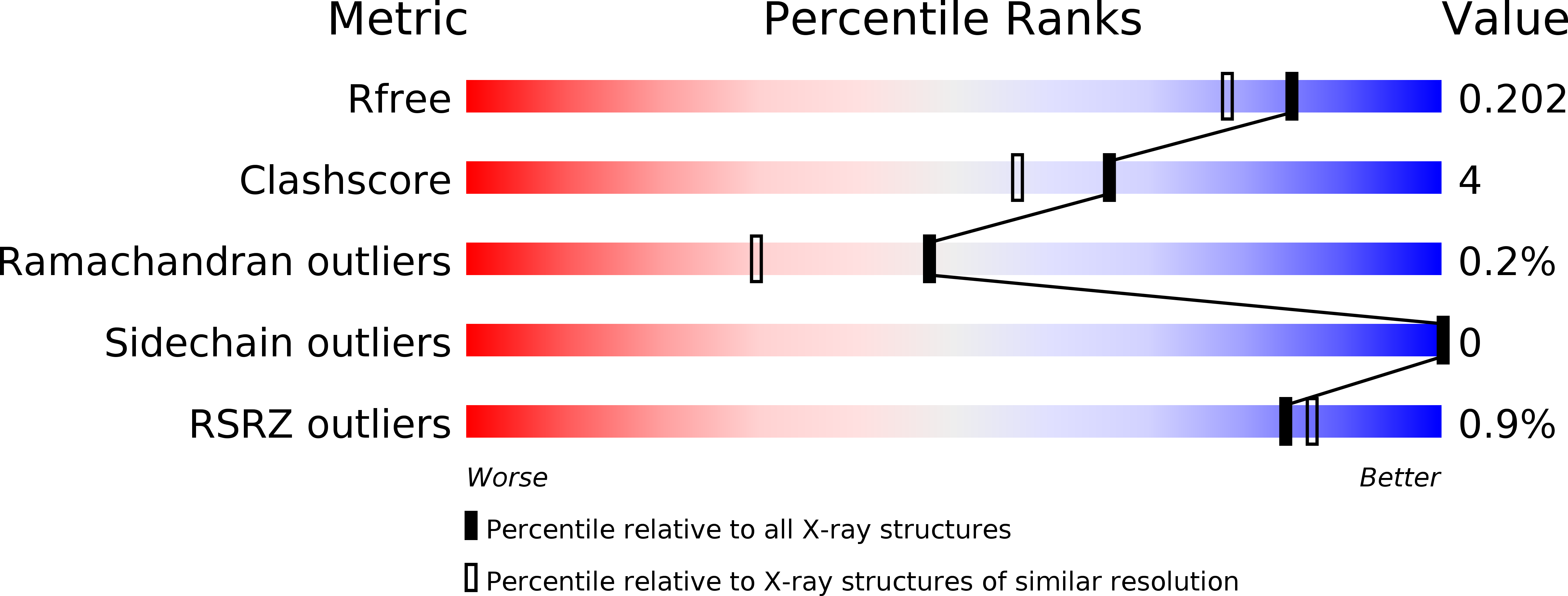
6N1E image
Deposition Date 2018-11-08
Release Date 2019-05-01
Last Version Date 2023-10-11
Entry Detail
PDB ID:
6N1E
Keywords:
Title:
Crystal structure of X. citri phosphoglucomutase in complex with 1-methyl-glucose 6-phosphate
Biological Source:
Source Organism:
Method Details:
Experimental Method:
Resolution:
1.70 Å
R-Value Free:
0.20
R-Value Work:
0.16
R-Value Observed:
0.16
Space Group:
P 21 21 21
Macromolecular Entities
Polymer Type:polypeptide(L)
Molecule:Phosphomannomutase/phosphoglucomutase
Chain IDs:A
Chain Length:468
Number of Molecules:1
Biological Source:Xanthomonas citri
Primary Citation
Synthesis, Derivatization, and Structural Analysis of Phosphorylated Mono-, Di-, and Trifluorinated d-Gluco-heptuloses by Glucokinase: Tunable Phosphoglucomutase Inhibition.
Acs Omega 4 7029 7037 (2019)
PMID: 31179410 DOI: 10.1021/acsomega.9b00008

Abstact

Glucokinase phosphorylated a series of C-1 fluorinated α-d-gluco-heptuloses. These phosphorylated products were discovered to be inhibitors of α-phosphomannomutase/phosphoglucomutase (αPMM/PGM) and β-phosphoglucomutase (βPGM). Inhibition potency with both mutases inversely correlated to the degree of fluorination. Structural analysis with αPMM demonstrated the inhibitor binding to the active site, with the phosphate in the phosphate binding site and the anomeric hydroxyl directed to the catalytic site.

Legend

Protein

Chemical

Disease

Primary Citation of related structures