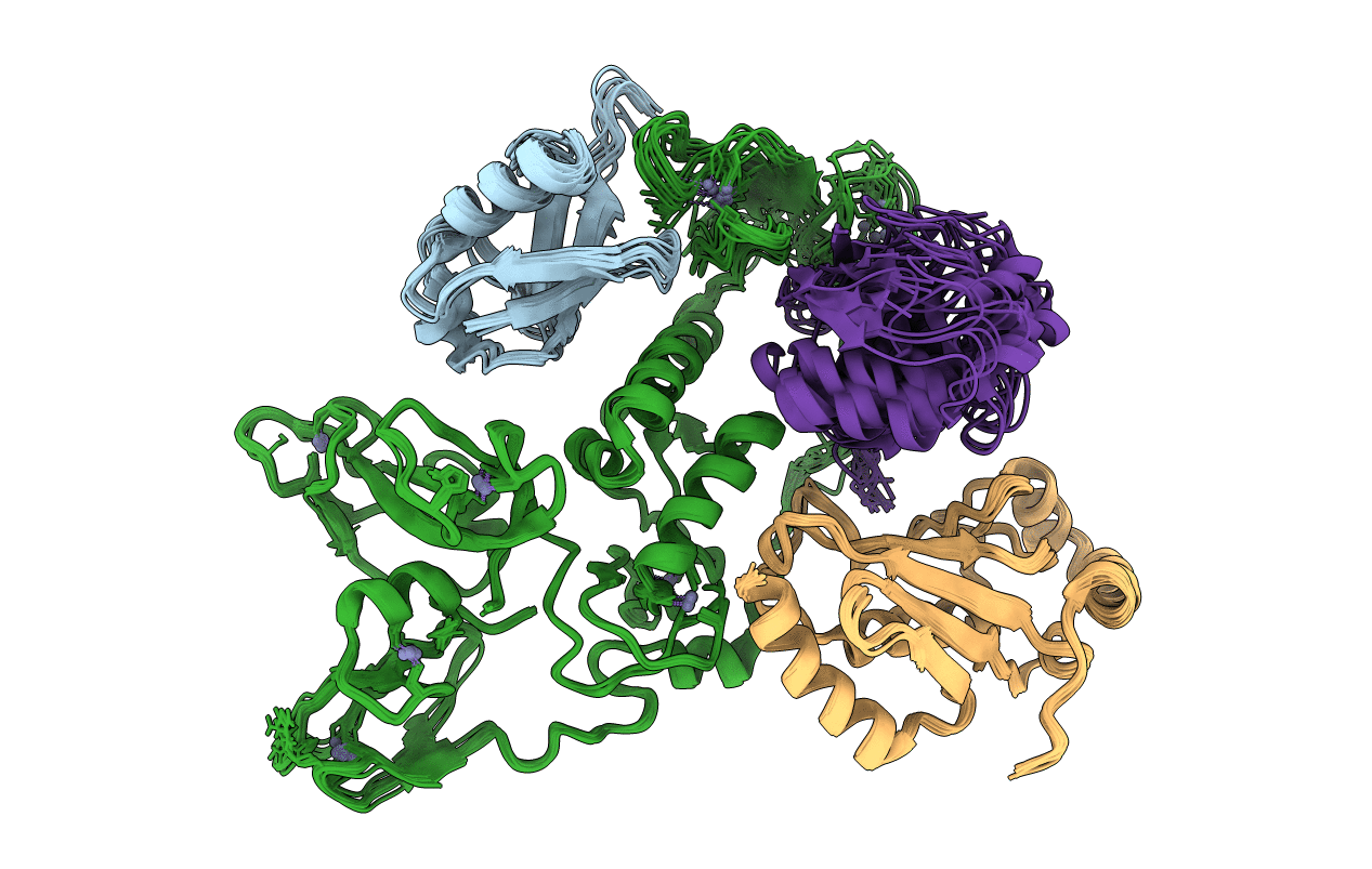
6N13 image
Entry Detail
PDB ID:
6N13
Keywords:
Title:
UbcH7-Ub Complex with R0RBR Parkin and phosphoubiquitin
Biological Source:
Source Organism:
Host Organism:
PDB Version:
Deposition Date:
2018-11-08
Release Date:
2018-11-28
Method Details:
Experimental Method:
Conformers Calculated:
1000
Conformers Submitted:
10
Selection Criteria:
structures with the lowest energy
Macromolecular Entities
Polymer Type:polypeptide(L)
Description:phosphoubiquitin
Chain IDs:D (auth: A)
Chain Length:76
Number of Molecules:1
Biological Source:Homo sapiens
Polymer Type:polypeptide(L)
Description:E3 ubiquitin-protein ligase parkin
Mutations:Q347C
Chain IDs:A (auth: B)
Chain Length:322
Number of Molecules:1
Biological Source:Homo sapiens
Polymer Type:polypeptide(L)
Description:Ubiquitin-conjugating enzyme E2 L3
Mutations:C17S, C86K, C137S
Chain IDs:C
Chain Length:156
Number of Molecules:1
Biological Source:Homo sapiens
Polymer Type:polypeptide(L)
Description:ubiquitin
Chain IDs:B (auth: D)
Chain Length:76
Number of Molecules:1
Biological Source:Homo sapiens
Modified Residue
Compound ID Chain ID Parent Comp ID Details 2D Image
SEP D SER modified residue
Ligand Molecules
Primary Citation
Synergistic recruitment of UbcH7~Ub and phosphorylated Ubl domain triggers parkin activation.
EMBO J. 37 ? ? (2018)
PMID: 30446597 DOI: 10.15252/embj.2018100014

Abstact

The E3 ligase parkin ubiquitinates outer mitochondrial membrane proteins during oxidative stress and is linked to early-onset Parkinson's disease. Parkin is autoinhibited but is activated by the kinase PINK1 that phosphorylates ubiquitin leading to parkin recruitment, and stimulates phosphorylation of parkin's N-terminal ubiquitin-like (pUbl) domain. How these events alter the structure of parkin to allow recruitment of an E2~Ub conjugate and enhanced ubiquitination is an unresolved question. We present a model of an E2~Ub conjugate bound to the phospho-ubiquitin-loaded C-terminus of parkin, derived from NMR chemical shift perturbation experiments. We show the UbcH7~Ub conjugate binds in the open state whereby conjugated ubiquitin binds to the RING1/IBR interface. Further, NMR and mass spectrometry experiments indicate the RING0/RING2 interface is re-modelled, remote from the E2 binding site, and this alters the reactivity of the RING2(Rcat) catalytic cysteine, needed for ubiquitin transfer. Our experiments provide evidence that parkin phosphorylation and E2~Ub recruitment act synergistically to enhance a weak interaction of the pUbl domain with the RING0 domain and rearrange the location of the RING2(Rcat) domain to drive parkin activity.

Legend

Protein

Chemical

Disease

Primary Citation of related structures