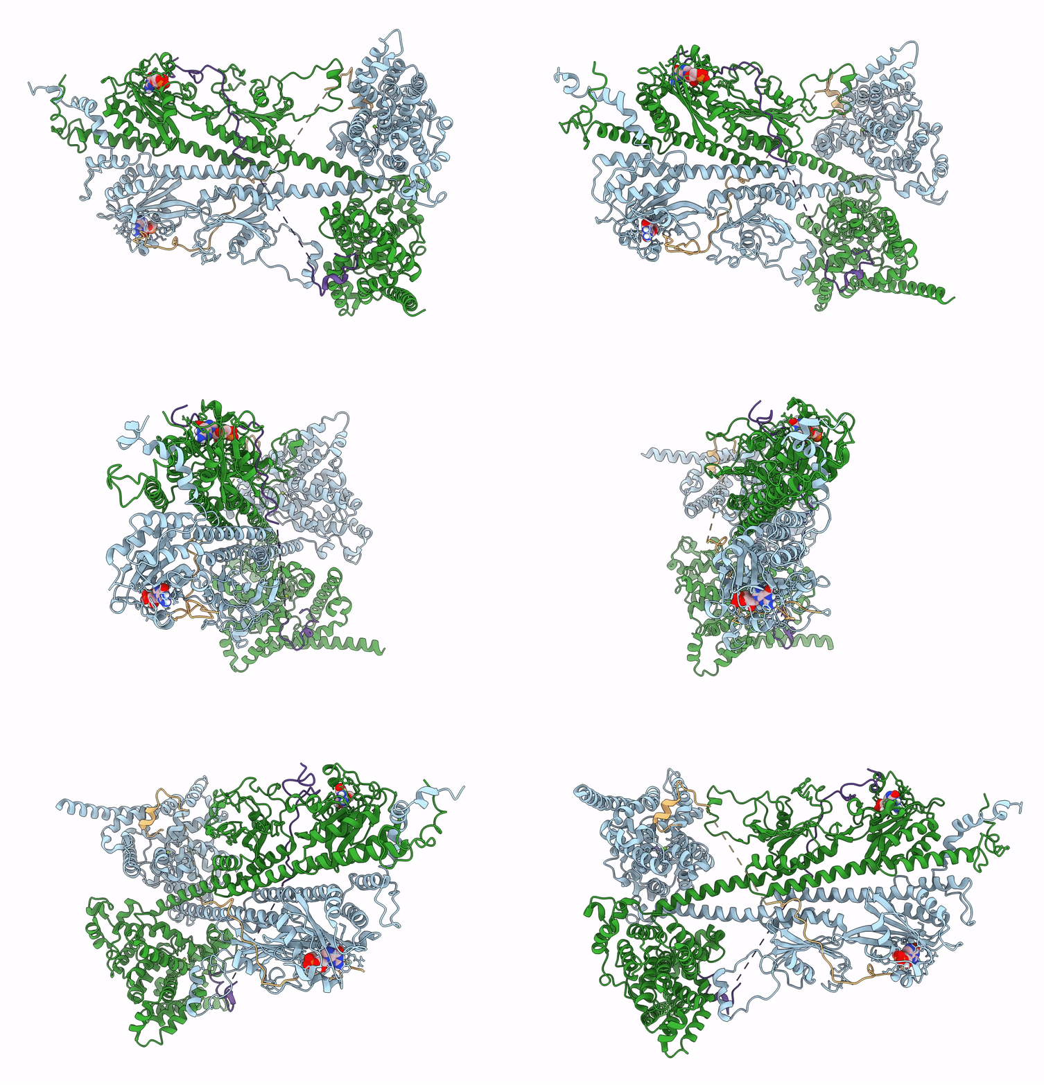
6MZB image
Deposition Date 2018-11-04
Release Date 2019-03-06
Last Version Date 2025-06-04
Entry Detail
PDB ID:
6MZB
Title:
Cryo-EM structure of phosphodiesterase 6
Biological Source:
Source Organism:
Bos taurus (Taxon ID: 9913)
Host Organism:
Method Details:
Experimental Method:
Resolution:
3.40 Å
Aggregation State:
PARTICLE
Reconstruction Method:
SINGLE PARTICLE
Macromolecular Entities
Polymer Type:polypeptide(L)
Molecule:Rod cGMP-specific 3',5'-cyclic phosphodiesterase subunit alpha
Gene (Uniprot):PDE6A
Chain IDs:B (auth: A)
Chain Length:859
Number of Molecules:1
Biological Source:Bos taurus
Polymer Type:polypeptide(L)
Molecule:Rod cGMP-specific 3',5'-cyclic phosphodiesterase subunit beta
Gene (Uniprot):PDE6B
Chain IDs:A (auth: B)
Chain Length:853
Number of Molecules:1
Biological Source:Bos taurus
Polymer Type:polypeptide(L)
Molecule:Retinal rod rhodopsin-sensitive cGMP 3',5'-cyclic phosphodiesterase subunit gamma
Gene (Uniprot):PDE6G
Chain IDs:C, D
Chain Length:87
Number of Molecules:2
Biological Source:Bos taurus
Primary Citation
Cryo-EM structure of phosphodiesterase 6 reveals insights into the allosteric regulation of type I phosphodiesterases.
Sci Adv 5 eaav4322 eaav4322 (2019)
PMID: 30820458 DOI: 10.1126/sciadv.aav4322

Abstact

Cyclic nucleotide phosphodiesterases (PDEs) work in conjunction with adenylate/guanylate cyclases to regulate the key second messengers of G protein-coupled receptor signaling. Previous attempts to determine the full-length structure of PDE family members at high-resolution have been hindered by structural flexibility, especially in their linker regions and N- and C-terminal ends. Therefore, most structure-activity relationship studies have so far focused on truncated and conserved catalytic domains rather than the regulatory domains that allosterically govern the activity of most PDEs. Here, we used single-particle cryo-electron microscopy to determine the structure of the full-length PDE6αβ2γ complex. The final density map resolved at 3.4 Å reveals several previously unseen structural features, including a coiled N-terminal domain and the interface of PDE6γ subunits with the PDE6αβ heterodimer. Comparison of the PDE6αβ2γ complex with the closed state of PDE2A sheds light on the conformational changes associated with the allosteric activation of type I PDEs.

Legend

Protein

Chemical

Disease

Primary Citation of related structures