Deposition Date
2018-11-02
Release Date
2019-11-06
Last Version Date
2023-10-11
Entry Detail
PDB ID:
6MYQ
Keywords:
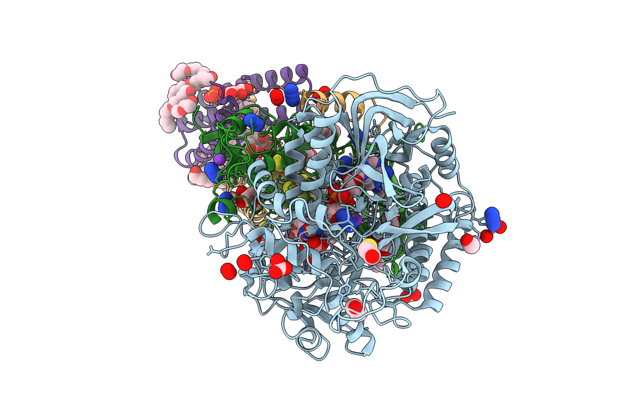
Title:
Avian mitochondrial complex II with ferulenol bound
Biological Source:
Source Organism(s):
Gallus gallus (Taxon ID: 9031)
Method Details:
Experimental Method:
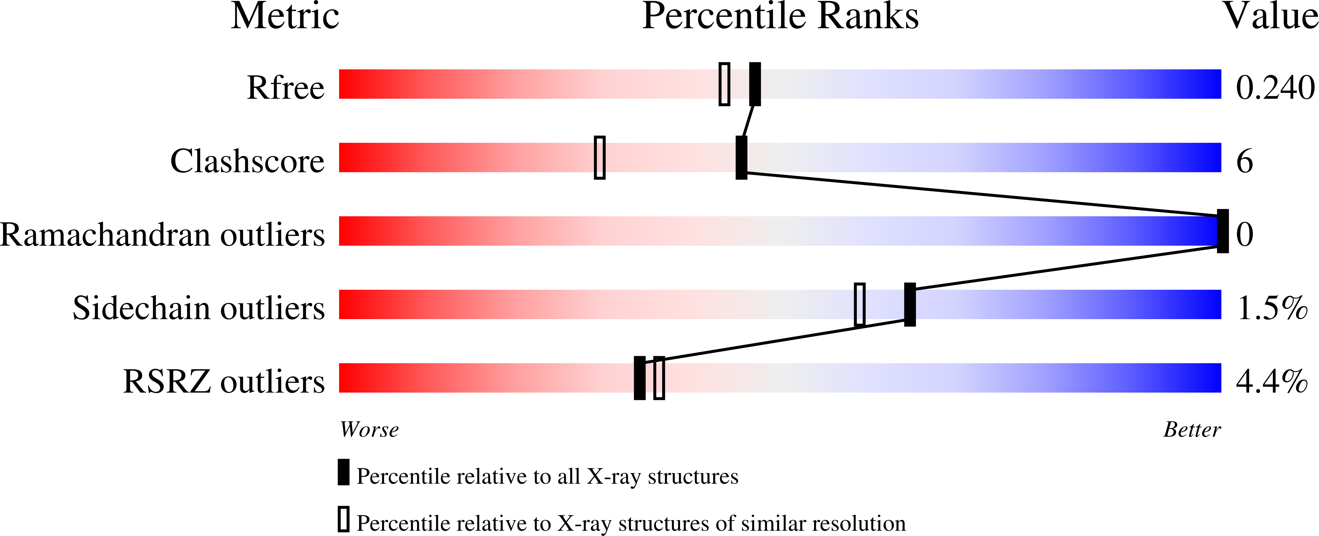
Resolution:
1.97 Å
R-Value Free:
0.23
R-Value Work:
0.20
R-Value Observed:
0.20
Space Group:
P 21 21 21