6MXB image
Entry Detail
PDB ID:
6MXB
Keywords:
Title:
Crystal structure of Trypanosoma brucei hypoxanthine-guanine-xanthine phosphoribosyltranferase in complex with XMP
Biological Source:
PDB Version:
Deposition Date:
2018-10-30
Release Date:
2019-07-24
Method Details:
Experimental Method:
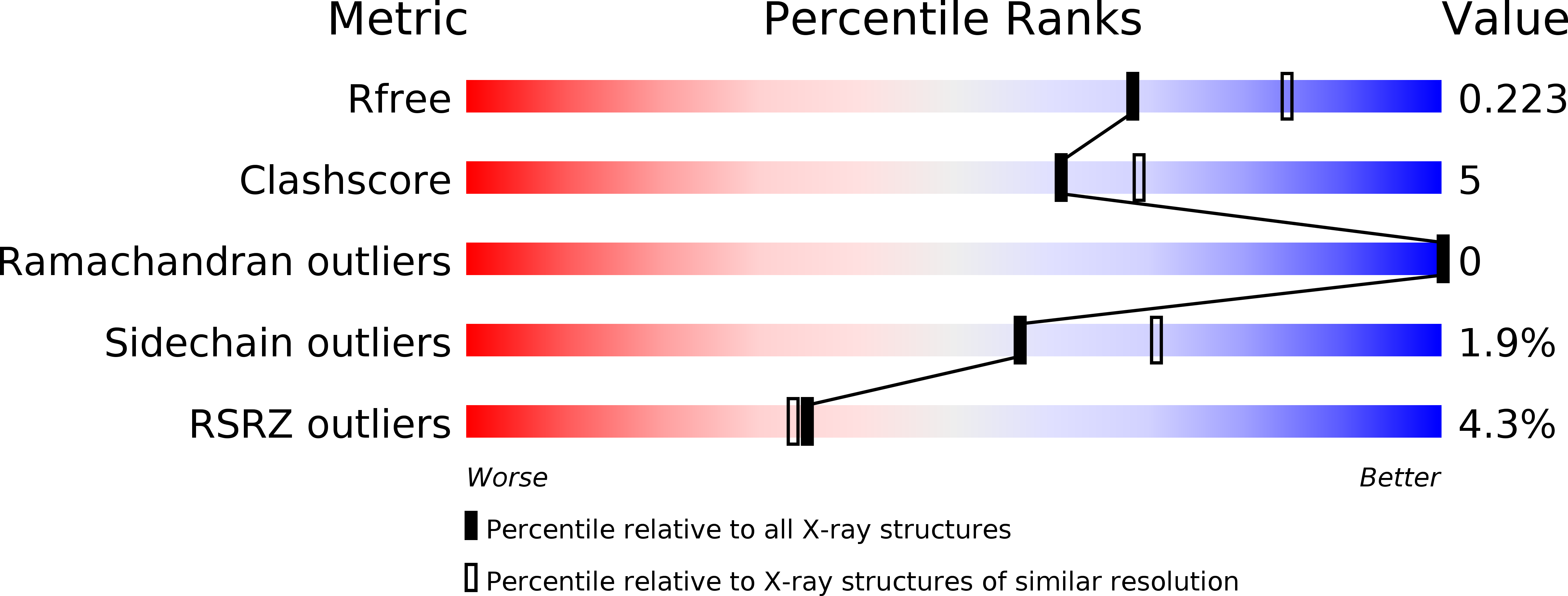
Resolution:
2.19 Å
R-Value Free:
0.22
R-Value Work:
0.18
R-Value Observed:
0.18
Space Group:
P 1 21 1
Macromolecular Entities
Polymer Type:polypeptide(L)
Description:Hypoxanthine-guanine phosphoribosyltransferase, putative
Chain IDs:A, B, C, D, E, F
Chain Length:272
Number of Molecules:6
Biological Source:Trypanosoma brucei brucei (strain 927/4 GUTat10.1)
Primary Citation
Crystal structures of Trypanosoma brucei hypoxanthine - guanine - xanthine phosphoribosyltransferase in complex with IMP, GMP and XMP.
Febs J. 286 4721 4736 (2019)
PMID: 31287615 DOI: 10.1111/febs.14987

Abstact

The 6-oxopurine phosphoribosyltransferases (PRTs) are drug targets for the treatment of parasitic diseases. This is due to the fact that parasites are auxotrophic for the 6-oxopurine bases relying on salvage enzymes for the synthesis of their 6-oxopurine nucleoside monophosphates. In Trypanosoma brucei, the parasite that is the aetiological agent for sleeping sickness, there are three 6-oxopurine PRT isoforms. Two are specific for hypoxanthine and guanine, whilst the third, characterized here, uses all three naturally occurring bases with similar efficiency. Here, we have determined crystal structures for TbrHGXPRT in complex with GMP, XMP and IMP to investigate the structural basis for substrate specificity. The results show that Y201 and E208, not commonly observed within the purine binding pocket of 6-oxopurine PRTs, contribute to the versatility of this enzyme. The structures further show that a nearby water can act as an adaptor to facilitate the binding of XMP and GMP. When GMP binds, a water can accept a proton from the 2-amino group but when XMP binds, the equivalent water can donate its proton to the 2-oxo group. However, when IMP is bound, no water molecule is observed at that location. DATABASE: Coordinates and structure factors were submitted to the Protein Data Bank and have accession codes of 6MXB, 6MXC, 6MXD and 6MXG for the TbrHGXPRT.XMP complex, TbrHGXPRT.GMP complex, TbrHGXPRT.IMP complex, and TbrHGPRT.XMP complex, respectively.

Legend

Protein

Chemical

Disease

Primary Citation of related structures