
Deposition Date
2018-10-08
Release Date
2018-12-12
Last Version Date
2024-10-09
Entry Detail
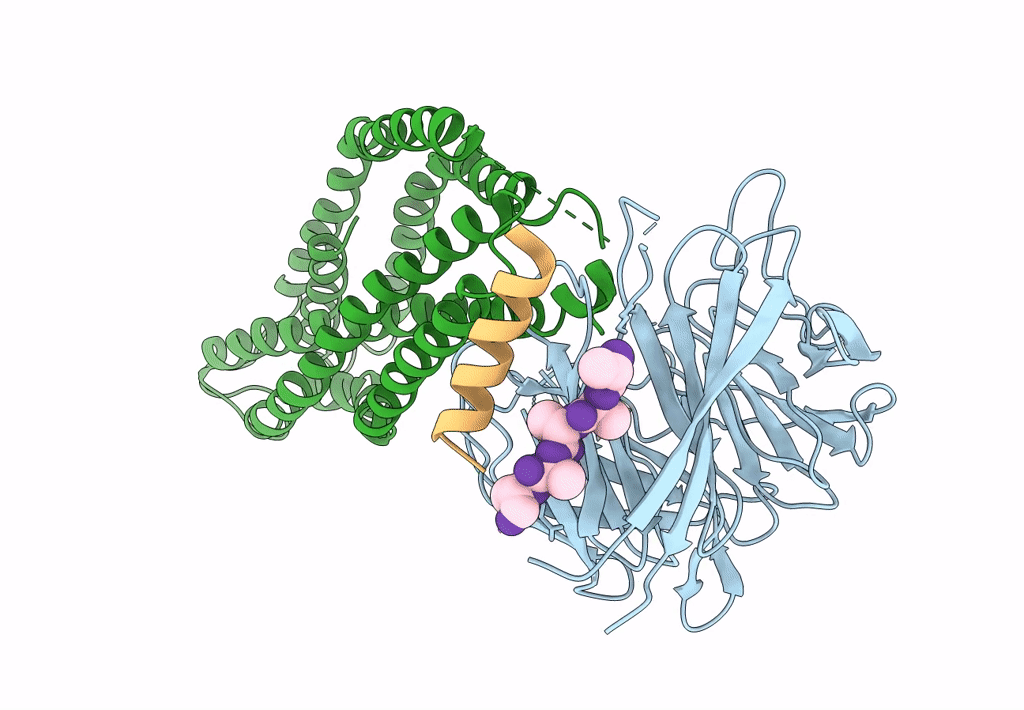
PDB ID:
6MPV
Keywords:
Title:
Cryo-electron microscopy structure of Plasmodium falciparum Rh5/CyRPA/Ripr invasion complex
Biological Source:
Source Organism(s):
Plasmodium falciparum (isolate 3D7) (Taxon ID: 36329)
Plasmodium falciparum (Taxon ID: 5833)
Plasmodium falciparum (Taxon ID: 5833)
Expression System(s):
Method Details:
Experimental Method:
Resolution:
7.17 Å
Aggregation State:
PARTICLE
Reconstruction Method:
SINGLE PARTICLE
