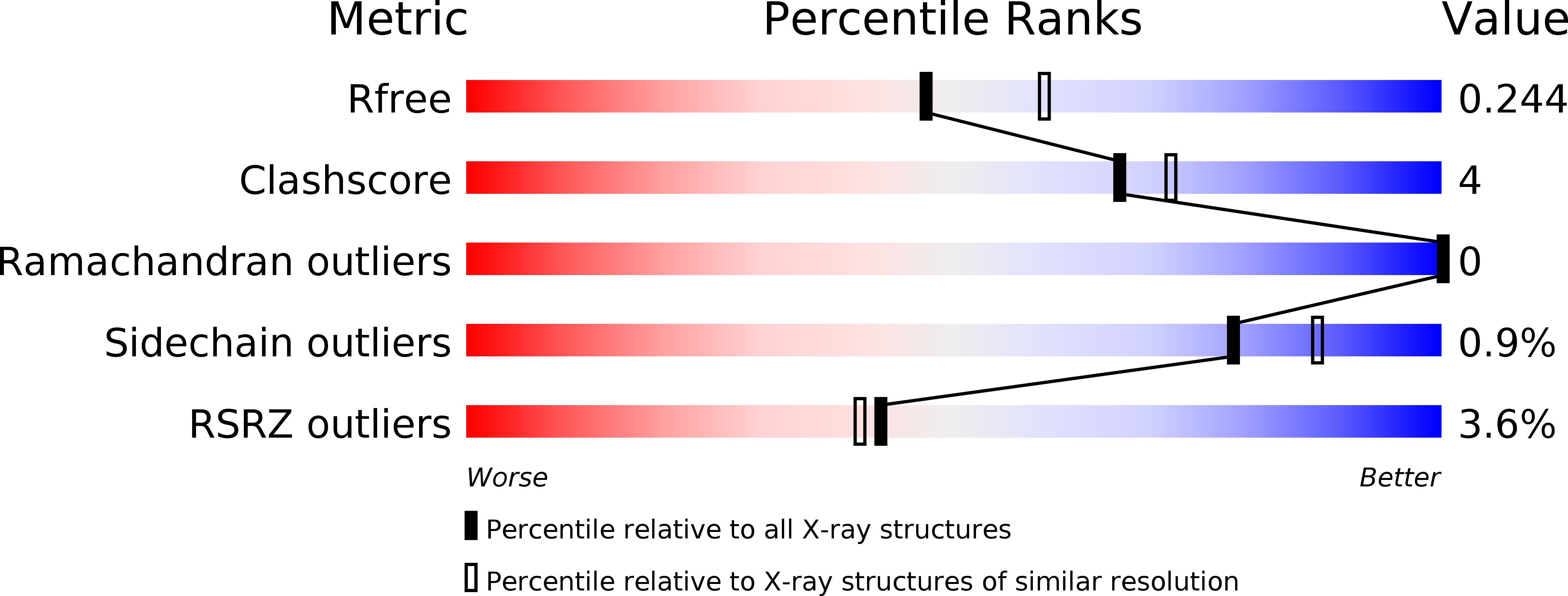
6MP1 image
Deposition Date 2018-10-05
Release Date 2018-11-07
Last Version Date 2024-11-20
Entry Detail
PDB ID:
6MP1
Keywords:
Title:
Crystal structures of the murine class I major histocompatibility complex H-2Db in complex with the mutant TRP1-K8 peptide
Biological Source:
Source Organism:
Mus musculus (Taxon ID: 10090)
Host Organism:
Method Details:
Experimental Method:
Resolution:
2.21 Å
R-Value Free:
0.24
R-Value Work:
0.20
R-Value Observed:
0.21
Space Group:
P 21 21 21
Macromolecular Entities
Polymer Type:polypeptide(L)
Molecule:TRP1-K8 peptide, Beta-2-microglobulin,H-2 class I histocompatibility antigen, D-B alpha chain, chimeric construct
Gene (Uniprot):B2m, H2-D1, Tyrp1
Mutations:Y84A, Y8K, A9M
Chain IDs:A
Chain Length:447
Number of Molecules:1
Biological Source:Mus musculus
Ligand Molecules
Primary Citation
Altered Binding of Tumor Antigenic Peptides to MHC Class I Affects CD8+T Cell-Effector Responses.
Cancer Immunol Res 6 1524 1536 (2018)
PMID: 30352798 DOI: 10.1158/2326-6066.CIR-18-0348

Abstact

T-cell priming occurs when a naïve T cell recognizes cognate peptide-MHC complexes on an activated antigen-presenting cell. The circumstances of this initial priming have ramifications on the fate of the newly primed T cell. Newly primed CD8+ T cells can embark onto different trajectories, with some becoming short-lived effector cells and others adopting a tissue resident or memory cell fate. To determine whether T-cell priming influences the quality of the effector T-cell response to tumors, we used transnuclear CD8+ T cells that recognize the melanoma antigen TRP1 using TRP1high or TRP1low TCRs that differ in both affinity and fine specificity. From a series of altered peptide ligands, we identified a point mutation (K8) in a nonanchor residue that, when analyzed crystallographically and biophysically, destabilized the peptide interaction with the MHC binding groove. In vitro, the K8 peptide induced robust proliferation of both TRP1high and TRP1low CD8+ T cells but did not induce expression of PD-1. Cytokine production from K8-stimulated TRP1 cells was minimal, whereas cytotoxicity was increased. Upon transfer into B16 tumor-bearing mice, the reference peptide (TRP1-M9)- and K8-stimulated TRP1 cells were equally effective at controlling tumor growth but accomplished this through different mechanisms. TRP1-M9-stimulated cells produced more IFNγ, whereas K8-stimulated cells accumulated to higher numbers and were more cytotoxic. We, therefore, conclude that TCR recognition of weakly binding peptides during priming can skew the effector function of tumor-specific CD8+ T cells.

Legend

Protein

Chemical

Disease

Primary Citation of related structures