Planned Maintenance: Some services may turn out to be unavailable from 15th January, 2026 to 16th January, 2026. We apologize for the inconvenience!

6MNI image
Deposition Date 2018-10-01
Release Date 2019-10-16
Last Version Date 2023-10-11
Entry Detail
PDB ID:
6MNI
Title:
Structure of the tandem CACHE domain of PscC
Biological Source:
Source Organism(s):
Expression System(s):
Method Details:
Experimental Method:
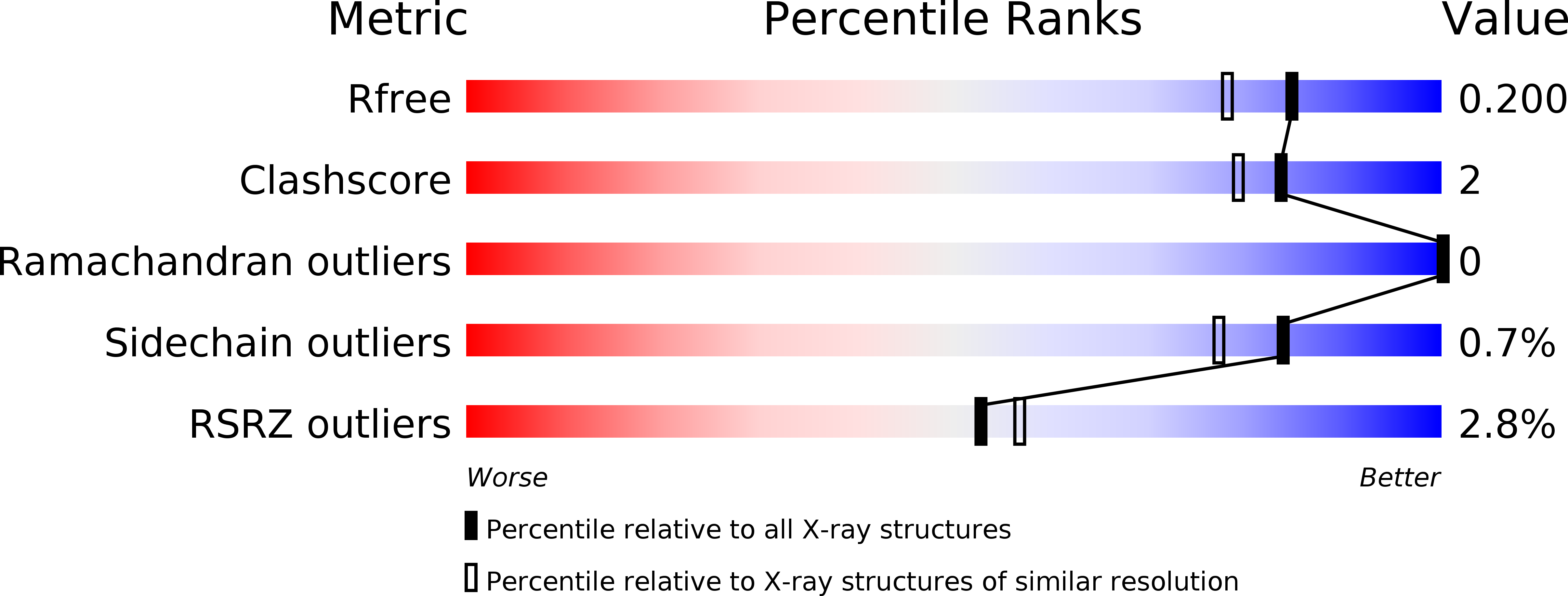
Resolution:
1.70 Å
R-Value Free:
0.19
R-Value Work:
0.16
R-Value Observed:
0.16
Space Group:
P 1
Macromolecular Entities
Polymer Type:polypeptide(L)
Molecule:Methyl-accepting chemotaxis protein
Chain IDs:A, B
Chain Length:287
Number of Molecules:2
Biological Source:Pseudomonas syringae pv. actinidiae
Primary Citation
Structure of a double CACHE chemoreceptor ligand-binding domain from Pseudomonas syringae provides insights into the basis of proline recognition.
Biochem.Biophys.Res.Commun. 549 194 199 (2021)
PMID: 33721671 DOI: 10.1016/j.bbrc.2021.02.090

Abstact

Chemotaxis is the process of sensing chemical gradients and navigating towards favourable conditions. Bacterial chemotaxis is mediated by arrays of trans-membrane chemoreceptor proteins. The most common class of chemoreceptors have periplasmic ligand-binding domains (LBDs) that detect extracellular chemical signs and transduce these signals to the downstream chemotaxis machinery. The repertoire of chemoreceptor proteins in a bacterium determines the range of environmental signals to which it can respond. Pseudomonas syringae pv. actinidiae (Psa) is a plant pathogen which causes bacterial canker of kiwifruit (Actinidia sp.). Compared to many other bacteria, Psa has a large number of chemoreceptors encoded in its genome (43) and most of these remain uncharacterized. A previous study identified PscC as a potential chemoreceptor for l-proline and other amino acid ligands. Here, we have characterized the interaction of PscC-LBD with l-proline using a combination of isothermal titration calorimetry (ITC) and X-ray crystallography. ITC confirmed direct binding of l-proline to PscC-LBD with KD value of 5.0 μM. We determined the structure of PscC-LBD in complex with l-proline. Our structural analysis showed that PscC-LBD adopts similar double-CACHE fold to several other amino acid chemoreceptors. A comparison of the PscC-LDB to other dCACHE structures highlights residues in the binding cavity which contribute to its ligand specificity.

Legend

Protein

Chemical

Disease

Primary Citation of related structures
Feedback Form
Name
Email
Institute
Feedback