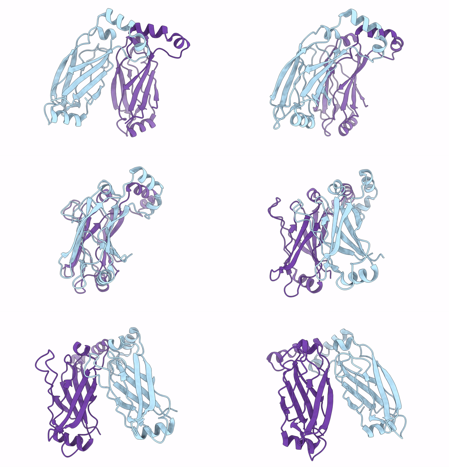
6MLU image
Deposition Date 2018-09-28
Release Date 2018-10-17
Last Version Date 2025-06-04
Entry Detail
PDB ID:
6MLU
Title:
Cryo-EM structure of lipid droplet formation protein Seipin/BSCL2
Biological Source:
Source Organism:
Host Organism:
Method Details:
Experimental Method:
Resolution:
4.00 Å
Aggregation State:
PARTICLE
Reconstruction Method:
SINGLE PARTICLE
Macromolecular Entities
Polymer Type:polypeptide(L)
Molecule:Seipin
Gene (Uniprot):Seipin
Chain IDs:A, B
Chain Length:382
Number of Molecules:2
Biological Source:Drosophila melanogaster
Ligand Molecules
Primary Citation
Cryo-electron microscopy structure of the lipid droplet-formation protein seipin.
J. Cell Biol. 217 4080 4091 (2018)
PMID: 30327422 DOI: 10.1083/jcb.201809067

Abstact

Metabolic energy is stored in cells primarily as triacylglycerols in lipid droplets (LDs), and LD dysregulation leads to metabolic diseases. The formation of monolayer-bound LDs from the endoplasmic reticulum (ER) bilayer is poorly understood, but the ER protein seipin is essential to this process. In this study, we report a cryo-electron microscopy structure and functional characterization of Drosophila melanogaster seipin. The structure reveals a ring-shaped dodecamer with the luminal domain of each monomer resolved at ∼4.0 Å. Each luminal domain monomer exhibits two distinctive features: a hydrophobic helix (HH) positioned toward the ER bilayer and a β-sandwich domain with structural similarity to lipid-binding proteins. This structure and our functional testing in cells suggest a model in which seipin oligomers initially detect forming LDs in the ER via HHs and subsequently act as membrane anchors to enable lipid transfer and LD growth.

Legend

Protein

Chemical

Disease

Primary Citation of related structures