6MLH image
Entry Detail
PDB ID:
6MLH
Keywords:
Title:
Crystal structure of X. citri phosphoglucomutase in complex with GLUCOPYRANOSYL-1-METHYL-PHOSPHONIC ACID
Biological Source:
Source Organism:
Host Organism:
PDB Version:
Deposition Date:
2018-09-27
Release Date:
2019-07-31
Method Details:
Experimental Method:
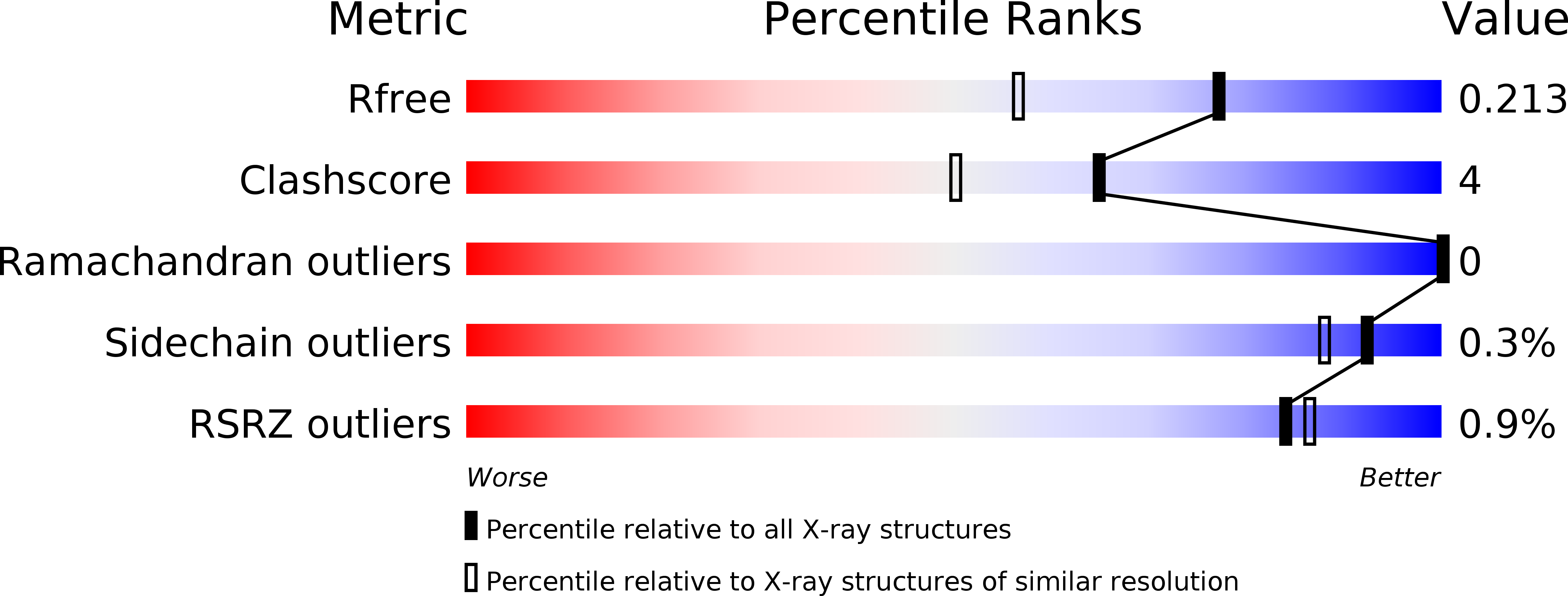
Resolution:
1.65 Å
R-Value Free:
0.21
R-Value Work:
0.17
R-Value Observed:
0.17
Space Group:
P 21 21 21
Macromolecular Entities
Polymer Type:polypeptide(L)
Description:Phosphoglucomutase
Chain IDs:A
Chain Length:468
Number of Molecules:1
Biological Source:Xanthomonas citri
Modified Residue
Compound ID Chain ID Parent Comp ID Details 2D Image
SEP A SER modified residue
Primary Citation
Inhibitory Evaluation of alpha PMM/PGM fromPseudomonas aeruginosa: Chemical Synthesis, Enzyme Kinetics, and Protein Crystallographic Study.
J.Org.Chem. 84 9627 9636 (2019)
PMID: 31264865 DOI: 10.1021/acs.joc.9b01305

Abstact

α-Phosphomannomutase/phosphoglucomutase (αPMM/PGM) from P. aeruginosa is involved in bacterial cell wall assembly and is implicated in P. aeruginosa virulence, yet few studies have addressed αPMM/PGM inhibition from this important Gram-negative bacterial human pathogen. Four structurally different α-d-glucopyranose 1-phosphate (αG1P) derivatives including 1-C-fluoromethylated analogues (1-3), 1,2-cyclic phosph(on)ate analogues (4-6), isosteric methylene phosphono analogues (7 and 8), and 6-fluoro-αG1P (9), were synthesized and assessed as potential time-dependent or reversible αPMM/PGM inhibitors. The resulting kinetic data were consistent with the crystallographic structures of the highly homologous Xanthomonas citri αPGM with inhibitors 3 and 7-9 binding to the enzyme active site (1.65-1.9 Å). These structural and kinetic insights will enhance the design of future αPMM/PGM inhibitors.

Legend

Protein

Chemical

Disease

Primary Citation of related structures