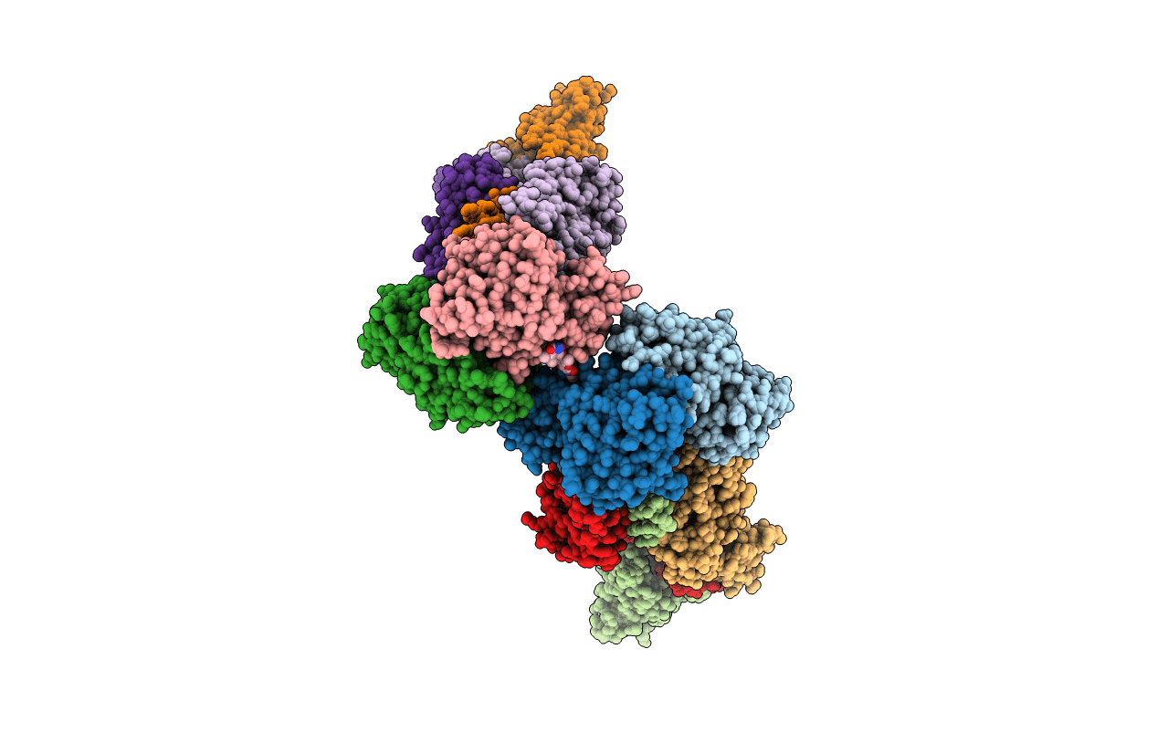
6MIT image
Entry Detail
PDB ID:
6MIT
Keywords:
Title:
LptBFGC from Enterobacter cloacae
Biological Source:
PDB Version:
Deposition Date:
2018-09-20
Release Date:
2019-03-27
Method Details:
Experimental Method:
Resolution:
3.20 Å
R-Value Free:
0.32
R-Value Work:
0.27
R-Value Observed:
0.27
Space Group:
P 21 2 21
Macromolecular Entities
Polymer Type:polypeptide(L)
Description:Lipopolysaccharide export system ATP-binding protein
Chain IDs:A, B, F (auth: D), G (auth: E)
Chain Length:241
Number of Molecules:4
Biological Source:Enterobacter cloacae subsp. cloacae (strain ATCC 13047 / DSM 30054 / NBRC 13535 / NCDC 279-56)
Polymer Type:polypeptide(L)
Description:Lipopolysaccharide export system protein LptC
Chain IDs:C, H
Chain Length:195
Number of Molecules:2
Biological Source:Enterobacter cloacae subsp. cloacae (strain ATCC 13047 / DSM 30054 / NBRC 13535 / NCDC 279-56)
Polymer Type:polypeptide(L)
Description:LPS export ABC transporter permease LptF
Chain IDs:D (auth: F), I
Chain Length:366
Number of Molecules:2
Biological Source:Enterobacter cloacae
Polymer Type:polypeptide(L)
Description:Lipopolysaccharide export system permease protein LptG
Chain IDs:E (auth: G), J
Chain Length:360
Number of Molecules:2
Biological Source:Enterobacter cloacae subsp. cloacae (strain ATCC 13047 / DSM 30054 / NBRC 13535 / NCDC 279-56)
Primary Citation
Structural basis of unidirectional export of lipopolysaccharide to the cell surface.
Nature 567 550 553 (2019)
PMID: 30894747 DOI: 10.1038/s41586-019-1039-0

Abstact

Gram-negative bacteria are surrounded by an inner cytoplasmic membrane and by an outer membrane, which serves as a protective barrier to limit entry of many antibiotics. The distinctive properties of the outer membrane are due to the presence of lipopolysaccharide1. This large glycolipid, which contains numerous sugars, is made in the cytoplasm; a complex of proteins forms a membrane-to-membrane bridge that mediates transport of lipopolysaccharide from the inner membrane to the cell surface1. The inner-membrane components of the protein bridge comprise an ATP-binding cassette transporter that powers transport, but how this transporter ensures unidirectional lipopolysaccharide movement across the bridge to the outer membrane is unknown2. Here we describe two crystal structures of a five-component inner-membrane complex that contains all the proteins required to extract lipopolysaccharide from the membrane and pass it to the protein bridge. Analysis of these structures, combined with biochemical and genetic experiments, identifies the path of lipopolysaccharide entry into the cavity of the transporter and up to the bridge. We also identify a protein gate that must open to allow movement of substrate from the cavity onto the bridge. Lipopolysaccharide entry into the cavity is ATP-independent, but ATP is required for lipopolysaccharide movement past the gate and onto the bridge. Our findings explain how the inner-membrane transport complex controls efficient unidirectional transport of lipopolysaccharide against its concentration gradient.

Legend

Protein

Chemical

Disease

Primary Citation of related structures