6MI0 image
Deposition Date 2018-09-18
Release Date 2019-07-10
Last Version Date 2023-10-11
Entry Detail
PDB ID:
6MI0
Keywords:
Title:
Crystal structure of the P450 domain of the CYP51-ferredoxin fusion protein from Methylococcus capsulatus, ligand-free state
Biological Source:
Method Details:
Experimental Method:
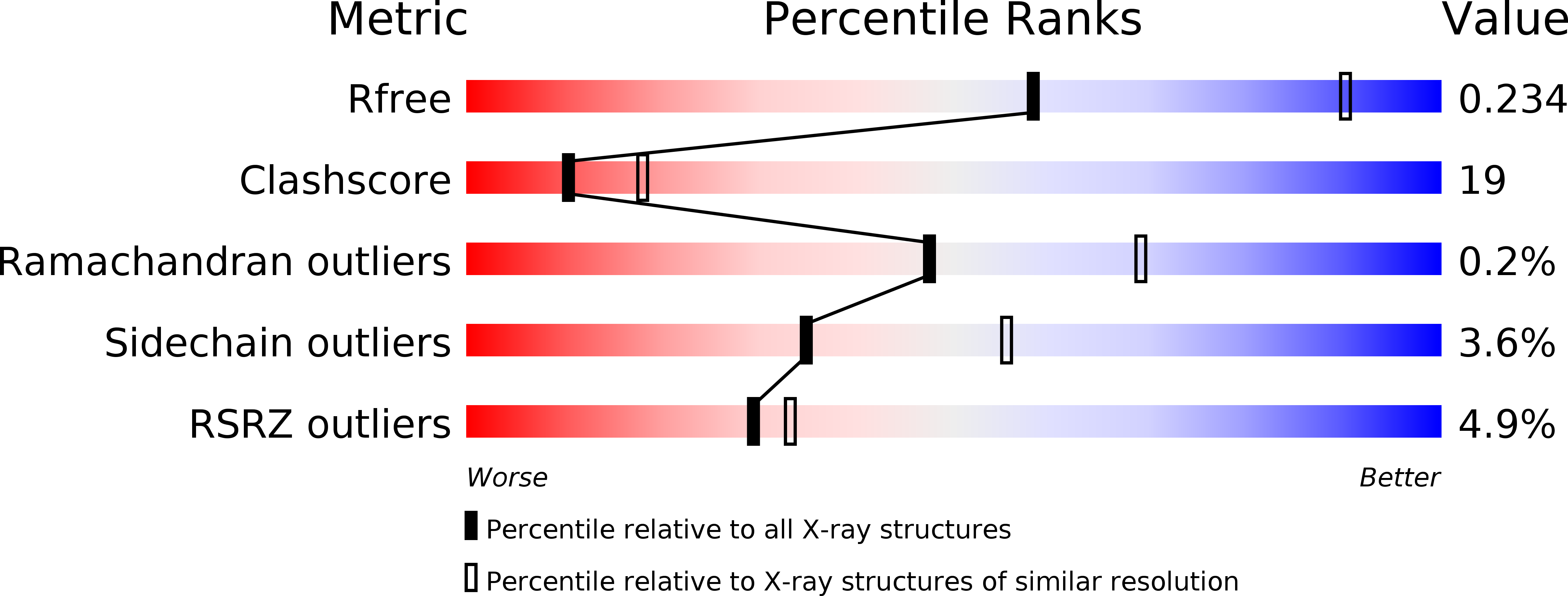
Resolution:
2.73 Å
R-Value Free:
0.23
R-Value Work:
0.21
R-Value Observed:
0.21
Space Group:
P 31 2 1
Macromolecular Entities
Structures with similar UniProt ID
Protein Blast
Polymer Type:polypeptide(L)
Molecule:Cytochrome P450 51
Gene (Uniprot):cyp51
Chain IDs:A
Chain Length:449
Number of Molecules:1
Biological Source:Methylococcus capsulatus (strain ATCC 33009 / NCIMB 11132 / Bath)
Ligand Molecules
Primary Citation
Concerning P450 evolution: Structural Analyses Support Bacterial Origin of Sterol 14 alpha-Demethylases.
Mol.Biol.Evol. ? ? ? (2020)
PMID: 33031537 DOI: 10.1093/molbev/msaa260

Abstact

Sterol biosynthesis, primarily associated with eukaryotic kingdoms of life, occurs as an abbreviated pathway in the bacterium Methylococcus capsulatus. Sterol 14α-demethylation is an essential step in this pathway and is catalyzed by cytochrome P450 51 (CYP51). In M. capsulatus, the enzyme consists of the P450 domain naturally fused to a ferredoxin domain at the C-terminus (CYP51fx). The structure of M. capsulatus CYP51fx was solved to 2.7 Å resolution and is the first structure of a bacterial sterol biosynthetic enzyme. The structure contained one P450 molecule per asymmetric unit with no electron density seen for ferredoxin. We connect this with the requirement of P450 substrate binding in order to activate productive ferredoxin binding. Further, the structure of the P450 domain with bound detergent (which replaced the substrate upon crystallization) was solved to 2.4 Å resolution. Comparison of these two structures to the CYP51s from human, fungi, and protozoa reveals strict conservation of the overall protein architecture. However, the structure of an "orphan" P450 from nonsterol-producing Mycobacterium tuberculosis that also has CYP51 activity reveals marked differences, suggesting that loss of function in vivo might have led to alterations in the structural constraints. Our results are consistent with the idea that eukaryotic and bacterial CYP51s evolved from a common cenancestor and that early eukaryotes may have recruited CYP51 from a bacterial source. The idea is supported by bioinformatic analysis, revealing the presence of CYP51 genes in >1,000 bacteria from nine different phyla, >50 of them being natural CYP51fx fusion proteins.

Legend

Protein

Chemical

Disease

Primary Citation of related structures
Feedback Form
Name
Email
Institute
Feedback