Deposition Date
2018-09-04
Release Date
2018-12-12
Last Version Date
2023-10-11
Entry Detail
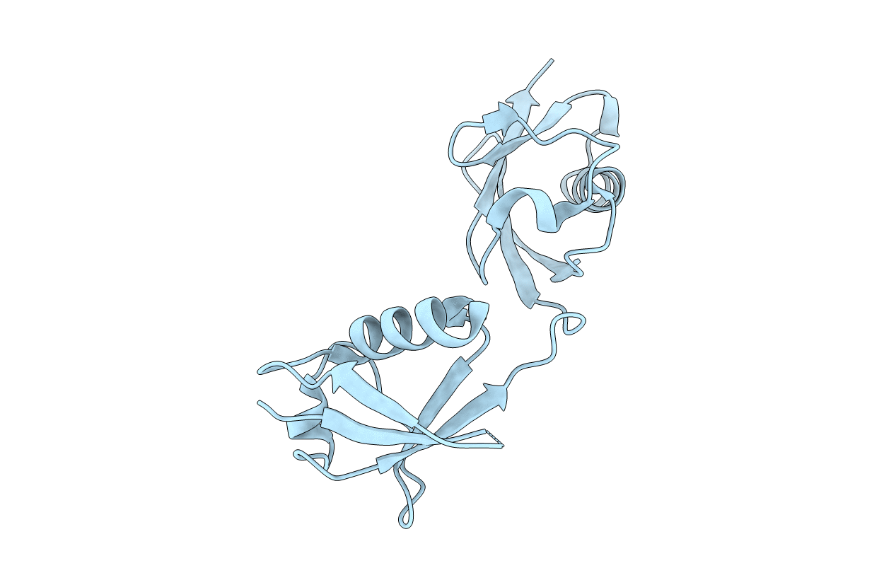
PDB ID:
6MDH
Keywords:
Title:
X-ray crystal structure of ISG15 from Myotis davidii
Biological Source:
Source Organism(s):
Myotis davidii (Taxon ID: 225400)
Expression System(s):
Method Details:
Experimental Method:
Resolution:
1.37 Å
R-Value Free:
0.19
R-Value Work:
0.16
R-Value Observed:
0.16
Space Group:
I 21 21 21