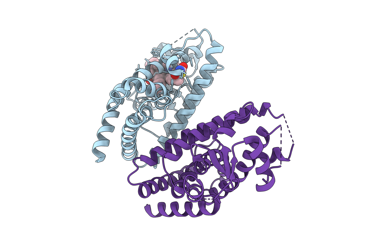
6MD4 image
Entry Detail
PDB ID:
6MD4
Title:
Crystal Structure of Human PPARgamma Ligand Binding Domain in Complex with Rosiglitazone and Oleic acid
Biological Source:
Source Organism:
PDB Version:
Deposition Date:
2018-09-03
Release Date:
2019-01-09
Method Details:
Experimental Method:
Resolution:
2.24 Å
R-Value Free:
0.28
R-Value Work:
0.24
R-Value Observed:
0.24
Space Group:
C 1 2 1
Macromolecular Entities
Polymer Type:polypeptide(L)
Description:Peroxisome proliferator-activated receptor gamma
Chain IDs:A, B
Chain Length:275
Number of Molecules:2
Biological Source:Homo sapiens
Primary Citation
Cooperative cobinding of synthetic and natural ligands to the nuclear receptor PPAR gamma.
Elife 7 ? ? (2018)
PMID: 30575522 DOI: 10.7554/eLife.43320

Abstact

Crystal structures of peroxisome proliferator-activated receptor gamma (PPARγ) have revealed overlapping binding modes for synthetic and natural/endogenous ligands, indicating competition for the orthosteric pocket. Here we show that cobinding of a synthetic ligand to the orthosteric pocket can push natural and endogenous PPARγ ligands (fatty acids) out of the orthosteric pocket towards an alternate ligand-binding site near the functionally important omega (Ω)-loop. X-ray crystallography, NMR spectroscopy, all-atom molecular dynamics simulations, and mutagenesis coupled to quantitative biochemical functional and cellular assays reveal that synthetic ligand and fatty acid cobinding can form a 'ligand link' to the Ω-loop and synergistically affect the structure and function of PPARγ. These findings contribute to a growing body of evidence indicating ligand binding to nuclear receptors can be more complex than the classical one-for-one orthosteric exchange of a natural or endogenous ligand with a synthetic ligand.

Legend

Protein

Chemical

Disease

Primary Citation of related structures