6MAB image
Deposition Date 2018-08-27
Release Date 2019-09-04
Last Version Date 2024-03-13
Entry Detail
PDB ID:
6MAB
Title:
1.7A resolution structure of RsbU from Chlamydia trachomatis (periplasmic domain)
Biological Source:
Source Organism:
Host Organism:
Method Details:
Experimental Method:
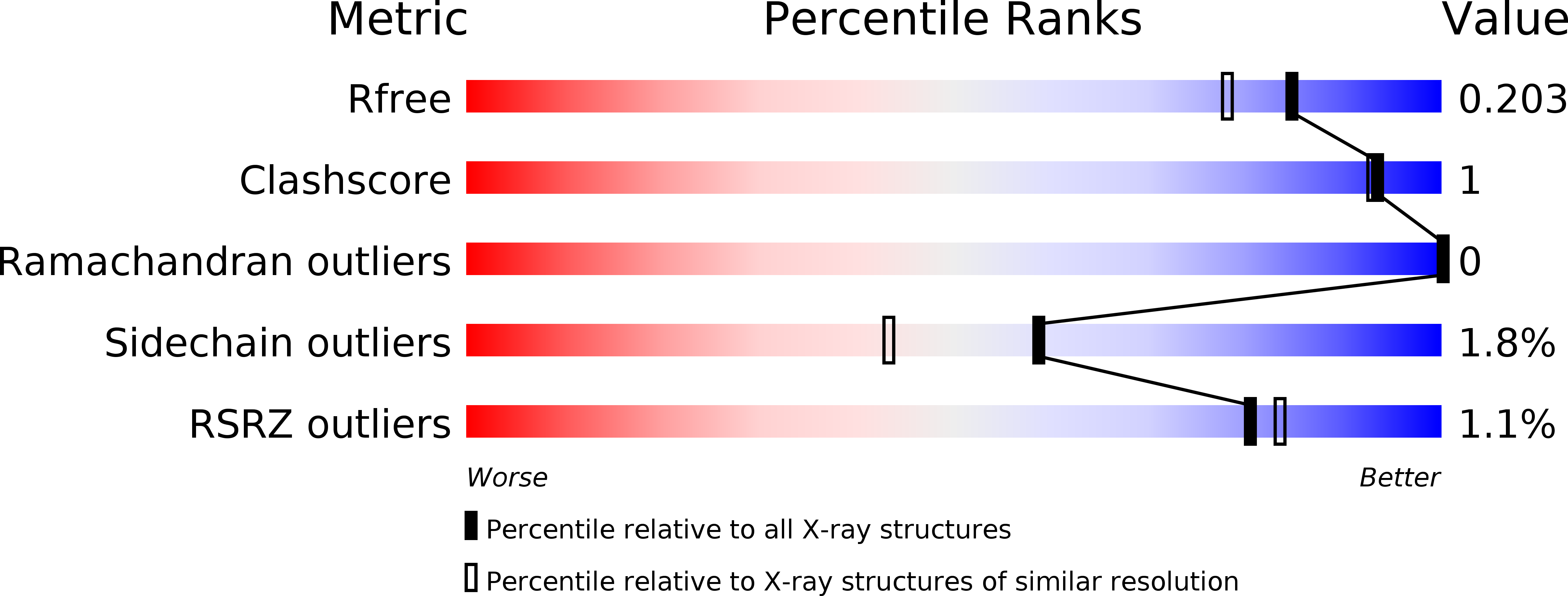
Resolution:
1.70 Å
R-Value Free:
0.19
R-Value Work:
0.16
R-Value Observed:
0.16
Space Group:
I 4
Macromolecular Entities
Structures with similar UniProt ID
Protein Blast
Polymer Type:polypeptide(L)
Molecule:Sigma regulatory family protein-PP2C phosphatase
Gene (Uniprot):rbsU
Chain IDs:A
Chain Length:272
Number of Molecules:1
Biological Source:Chlamydia trachomatis serovar L2
Ligand Molecules
Primary Citation
Structural and ligand binding analyses of the periplasmic sensor domain of RsbU in Chlamydia trachomatis support a role in TCA cycle regulation.
Mol.Microbiol. 113 68 88 (2020)
PMID: 31637787 DOI: 10.1111/mmi.14401

Abstact

Chlamydia trachomatis is an obligate intracellular bacteria that undergo dynamic morphologic and physiologic conversions upon gaining an access to a eukaryotic cell. These conversions likely require the detection of key environmental conditions and regulation of metabolic activity. Chlamydia encodes homologs to proteins in the Rsb phosphoregulatory partner-switching pathway, best described in Bacillus subtilis. ORF CT588 has a strong sequence similarity to RsbU cytoplasmic phosphatase domain but also contains a unique periplasmic sensor domain that is expected to control the phosphatase activity. A 1.7 Å crystal structure of the periplasmic domain of the RsbU protein from C. trachomatis (PDB 6MAB) displays close structural similarity to DctB from Vibrio and Sinorhizobium. DctB has been shown, both structurally and functionally, to specifically bind to the tricarboxylic acid (TCA) cycle intermediate succinate. Surface plasmon resonance and differential scanning fluorimetry of TCA intermediates and potential metabolites from a virtual screen of RsbU revealed that alpha-ketoglutarate, malate and oxaloacetate bound to the RsbU periplasmic domain. Substitutions in the putative binding site resulted in reduced binding capabilities. An RsbU null mutant showed severe growth defects which could be restored through genetic complementation. Chemical inhibition of ATP synthesis by oxidative phosphorylation phenocopied the growth defect observed in the RsbU null strain. Altogether, these data support a model with the Rsb system responding differentially to TCA cycle intermediates to regulate metabolism and key differentiation processes.

Legend

Protein

Chemical

Disease

Primary Citation of related structures