6M8R image
Deposition Date 2018-08-22
Release Date 2019-02-27
Last Version Date 2023-10-11
Entry Detail
PDB ID:
6M8R
Keywords:
Title:
Crystal structure of the KCTD16 BTB domain in complex with GABAB2 peptide
Biological Source:
Source Organism(s):
Homo sapiens (Taxon ID: 9606)
Method Details:
Experimental Method:
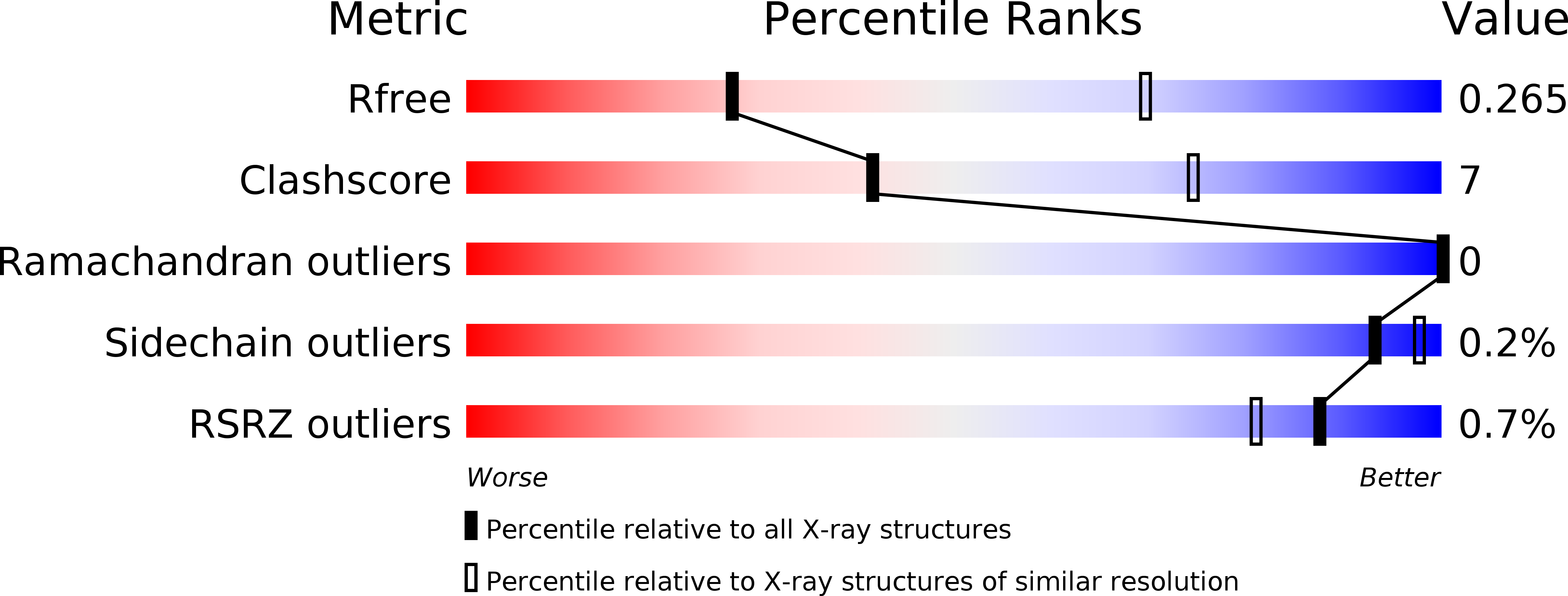
Resolution:
3.20 Å
R-Value Free:
0.26
R-Value Work:
0.21
R-Value Observed:
0.22
Space Group:
P 1 21 1
Macromolecular Entities
Structures with similar UniProt ID
Protein Blast
Polymer Type:polypeptide(L)
Molecule:BTB/POZ domain-containing protein KCTD16
Gene (Uniprot):KCTD16
Chain IDs:A (auth: G), B (auth: H), C (auth: I), D (auth: J), E (auth: B), F (auth: C), G (auth: D), H (auth: E), I (auth: A), J (auth: F)
Chain Length:103
Number of Molecules:10
Biological Source:Homo sapiens
Structures with similar UniProt ID
Protein Blast
Polymer Type:polypeptide(L)
Molecule:Gamma-aminobutyric acid type B receptor subunit 2
Gene (Uniprot):GABBR2
Chain IDs:K, L
Chain Length:41
Number of Molecules:2
Biological Source:Homo sapiens
Ligand Molecules
Primary Citation
Structural basis for KCTD-mediated rapid desensitization of GABABsignalling.
Nature 567 127 131 (2019)
PMID: 30814734 DOI: 10.1038/s41586-019-0990-0

Abstact

The GABAB (γ-aminobutyric acid type B) receptor is one of the principal inhibitory neurotransmitter receptors in the brain, and it signals through heterotrimeric G proteins to activate a variety of effectors, including G-protein-coupled inwardly rectifying potassium channels (GIRKs)1,2. GABAB-receptor signalling is tightly regulated by auxiliary subunits called KCTDs, which control the kinetics of GIRK activation and desensitization3-5. However, the mechanistic basis for KCTD modulation of GABAB signalling remains incompletely understood. Here, using a combination of X-ray crystallography, electron microscopy, and functional and biochemical experiments, we reveal the molecular details of KCTD binding to both GABAB receptors and G-protein βγ subunits. KCTDs associate with the receptor by forming an asymmetric pentameric ring around a region of the receptor carboxy-terminal tail, while a second KCTD domain, H1, engages in a symmetric interaction with five copies of Gβγ in which the G-protein subunits also interact directly with one another. We further show that KCTD binding to Gβγ is highly cooperative, defining a model in which KCTD proteins cooperatively strip G proteins from GIRK channels to induce rapid desensitization following receptor activation. These results provide a framework for understanding the molecular basis for the precise temporal control of GABAB signalling by KCTD proteins.

Legend

Protein

Chemical

Disease

Primary Citation of related structures
Feedback Form
Name
Email
Institute
Feedback