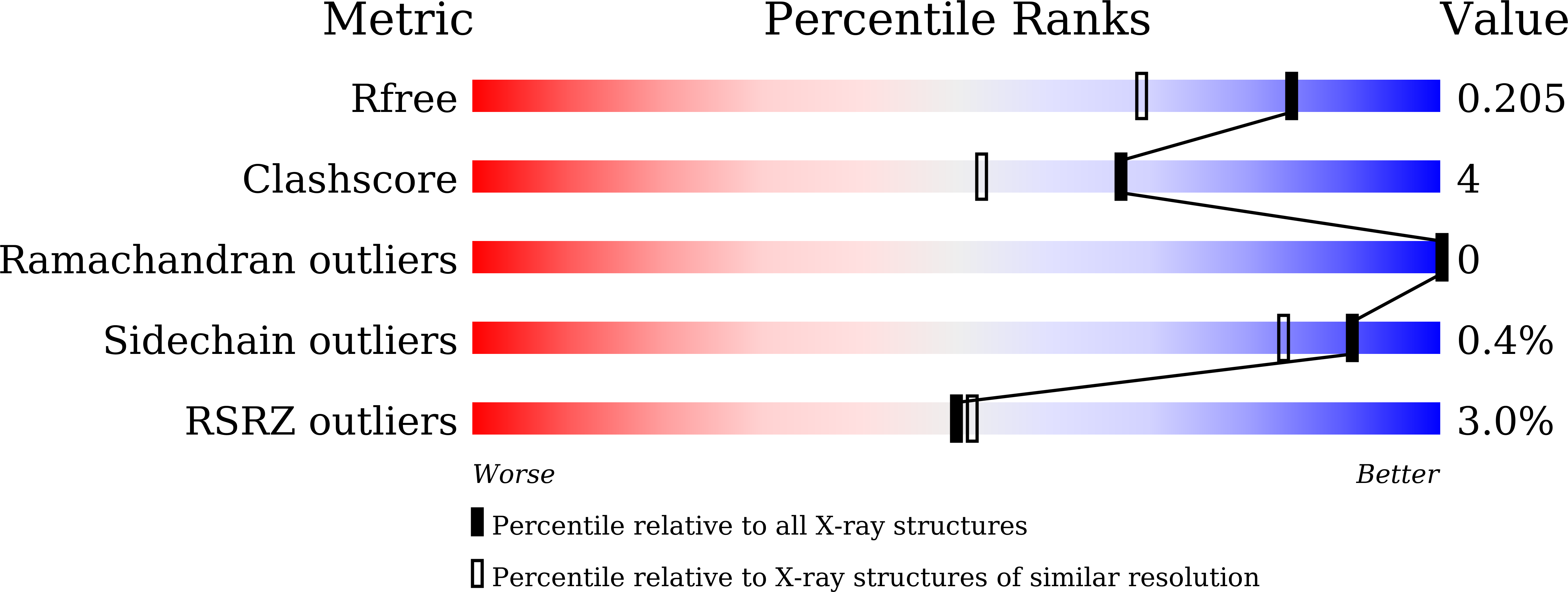
6M3G image
Deposition Date 2020-03-03
Release Date 2021-03-03
Last Version Date 2024-03-27
Entry Detail
PDB ID:
6M3G
Keywords:
Title:
Crystal structure of human HPF1
Biological Source:
Source Organism:
Homo sapiens (Taxon ID: 9606)
Method Details:
Experimental Method:
Resolution:
1.57 Å
R-Value Free:
0.20
R-Value Work:
0.18
R-Value Observed:
0.18
Space Group:
H 3 2
Macromolecular Entities
Polymer Type:polypeptide(L)
Molecule:Histone PARylation factor 1
Gene (Uniprot):HPF1
Chain IDs:A
Chain Length:346
Number of Molecules:1
Biological Source:Homo sapiens
Primary Citation
HPF1 remodels the active site of PARP1 to enable the serine ADP-ribosylation of histones.
Nat Commun 12 1028 1028 (2021)
PMID: 33589610 DOI: 10.1038/s41467-021-21302-4

Abstact

Upon binding to DNA breaks, poly(ADP-ribose) polymerase 1 (PARP1) ADP-ribosylates itself and other factors to initiate DNA repair. Serine is the major residue for ADP-ribosylation upon DNA damage, which strictly depends on HPF1. Here, we report the crystal structures of human HPF1/PARP1-CAT ΔHD complex at 1.98 Å resolution, and mouse and human HPF1 at 1.71 Å and 1.57 Å resolution, respectively. Our structures and mutagenesis data confirm that the structural insights obtained in a recent HPF1/PARP2 study by Suskiewicz et al. apply to PARP1. Moreover, we quantitatively characterize the key residues necessary for HPF1/PARP1 binding. Our data show that through salt-bridging to Glu284/Asp286, Arg239 positions Glu284 to catalyze serine ADP-ribosylation, maintains the local conformation of HPF1 to limit PARP1 automodification, and facilitates HPF1/PARP1 binding by neutralizing the negative charge of Glu284. These findings, along with the high-resolution structural data, may facilitate drug discovery targeting PARP1.

Legend

Protein

Chemical

Disease

Primary Citation of related structures