Deposition Date
2020-02-27
Release Date
2020-07-08
Last Version Date
2024-10-16
Entry Detail
PDB ID:
6M2J
Keywords:
Title:
Uncommon structural features of rabbit MHC class I (RLA-A1) complexed with rabbit haemorrhagic disease virus (RHDV) derived peptide, VP60-1
Biological Source:
Source Organism(s):
Oryctolagus cuniculus (Taxon ID: 9986)
Homo sapiens (Taxon ID: 9606)
Rabbit hemorrhagic disease virus (Taxon ID: 11976)
Homo sapiens (Taxon ID: 9606)
Rabbit hemorrhagic disease virus (Taxon ID: 11976)
Expression System(s):
Method Details:
Experimental Method:
Resolution:
2.20 Å
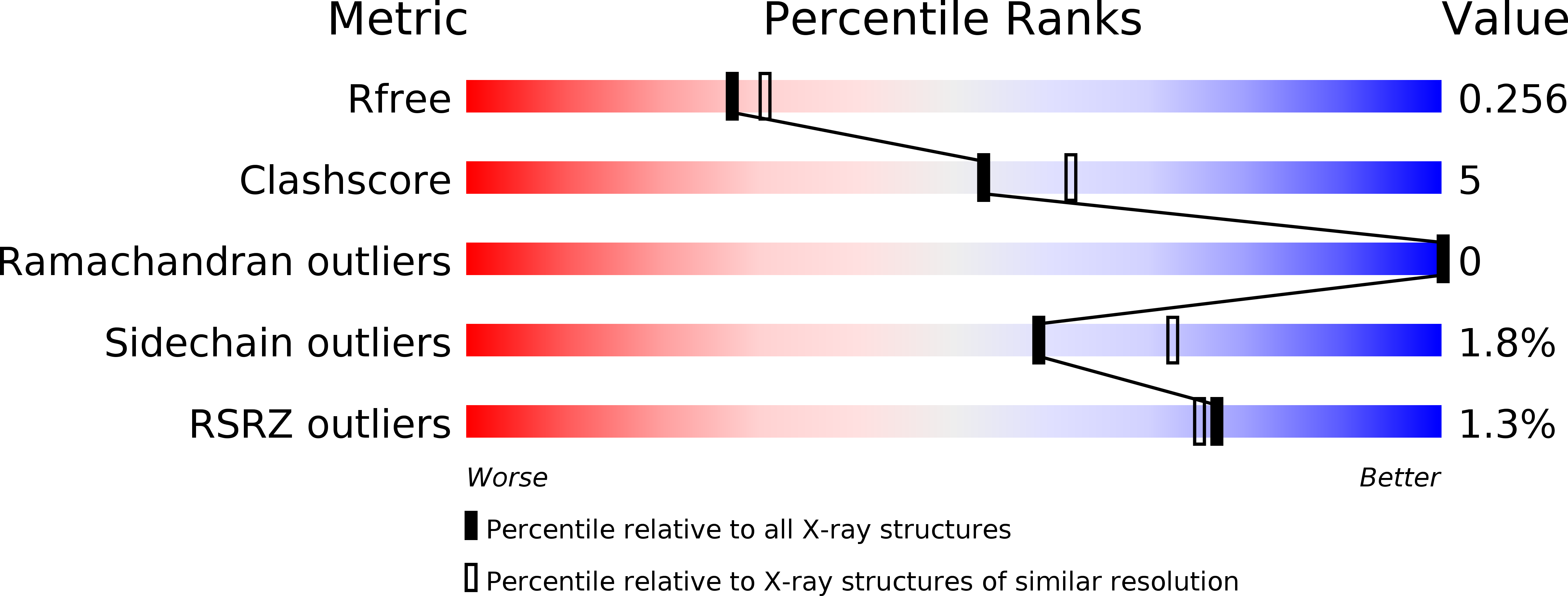
R-Value Free:
0.25
R-Value Work:
0.20
R-Value Observed:
0.20
Space Group:
P 21 21 21