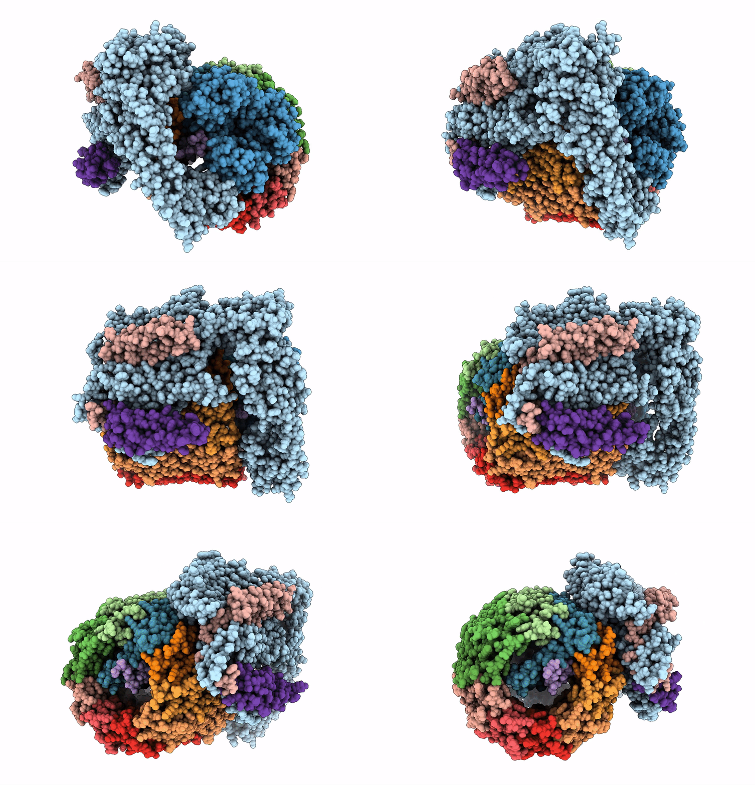
6M0S image
Entry Detail
PDB ID:
6M0S
EMDB ID:
Title:
3.6A Yeast Vo state3 prime
Biological Source:
PDB Version:
Deposition Date:
2020-02-22
Release Date:
2020-11-04
Method Details:
Experimental Method:
Resolution:
3.60 Å
Aggregation State:
PARTICLE
Reconstruction Method:
SINGLE PARTICLE
Macromolecular Entities
Polymer Type:polypeptide(L)
Description:V-type proton ATPase subunit a, vacuolar isoform
Chain IDs:A
Chain Length:825
Number of Molecules:1
Biological Source:Saccharomyces cerevisiae (strain ATCC 204508 / S288c)
Polymer Type:polypeptide(L)
Description:V-type proton ATPase subunit d
Chain IDs:D (auth: B)
Chain Length:345
Number of Molecules:1
Biological Source:Saccharomyces cerevisiae (strain ATCC 204508 / S288c)
Polymer Type:polypeptide(L)
Description:V-type proton ATPase subunit c''
Chain IDs:E (auth: C)
Chain Length:198
Number of Molecules:1
Biological Source:Saccharomyces cerevisiae (strain ATCC 204508 / S288c)
Polymer Type:polypeptide(L)
Description:V-type proton ATPase subunit c'
Chain IDs:F (auth: D)
Chain Length:158
Number of Molecules:1
Biological Source:Saccharomyces cerevisiae (strain ATCC 204508 / S288c)
Polymer Type:polypeptide(L)
Description:V-type proton ATPase subunit c
Chain IDs:G (auth: E), H (auth: F), I (auth: G), J (auth: H), K (auth: I), L (auth: J), M (auth: K), N (auth: L)
Chain Length:159
Number of Molecules:8
Biological Source:Saccharomyces cerevisiae (strain ATCC 204508 / S288c)
Polymer Type:polypeptide(L)
Description:V-type proton ATPase subunit e
Chain IDs:B (auth: M)
Chain Length:71
Number of Molecules:1
Biological Source:Saccharomyces cerevisiae (strain ATCC 204508 / S288c)
Polymer Type:polypeptide(L)
Description:V0 assembly protein 1
Chain IDs:O (auth: N)
Chain Length:52
Number of Molecules:1
Biological Source:Saccharomyces cerevisiae (strain ATCC 204508 / S288c)
Polymer Type:polypeptide(L)
Description:Uncharacterized protein YPR170W-B
Chain IDs:C (auth: O)
Chain Length:69
Number of Molecules:1
Biological Source:Saccharomyces cerevisiae (strain ATCC 204508 / S288c)
Ligand Molecules
Primary Citation
Cryo-EM and MD infer water-mediated proton transport and autoinhibition mechanisms of V o complex.
Sci Adv 6 ? ? (2020)
PMID: 33028525 DOI: 10.1126/sciadv.abb9605

Abstact

Rotary vacuolar adenosine triphosphatases (V-ATPases) drive transmembrane proton transport through a Vo proton channel subcomplex. Despite recent high-resolution structures of several rotary ATPases, the dynamic mechanism of proton pumping remains elusive. Here, we determined a 2.7-Å cryo-electron microscopy (cryo-EM) structure of yeast Vo proton channel in nanodisc that reveals the location of ordered water molecules along the proton path, details of specific protein-lipid interactions, and the architecture of the membrane scaffold protein. Moreover, we uncover a state of Vo that shows the c-ring rotated by ~14°. Molecular dynamics simulations demonstrate that the two rotary states are in thermal equilibrium and depict how the protonation state of essential glutamic acid residues couples water-mediated proton transfer with c-ring rotation. Our cryo-EM models and simulations also rationalize a mechanism for inhibition of passive proton transport as observed for free Vo that is generated as a result of V-ATPase regulation by reversible disassembly in vivo.

Legend

Protein

Chemical

Disease

Primary Citation of related structures