6LUO image
Deposition Date 2020-01-30
Release Date 2021-04-28
Last Version Date 2024-11-13
Entry Detail
PDB ID:
6LUO
Keywords:
Title:
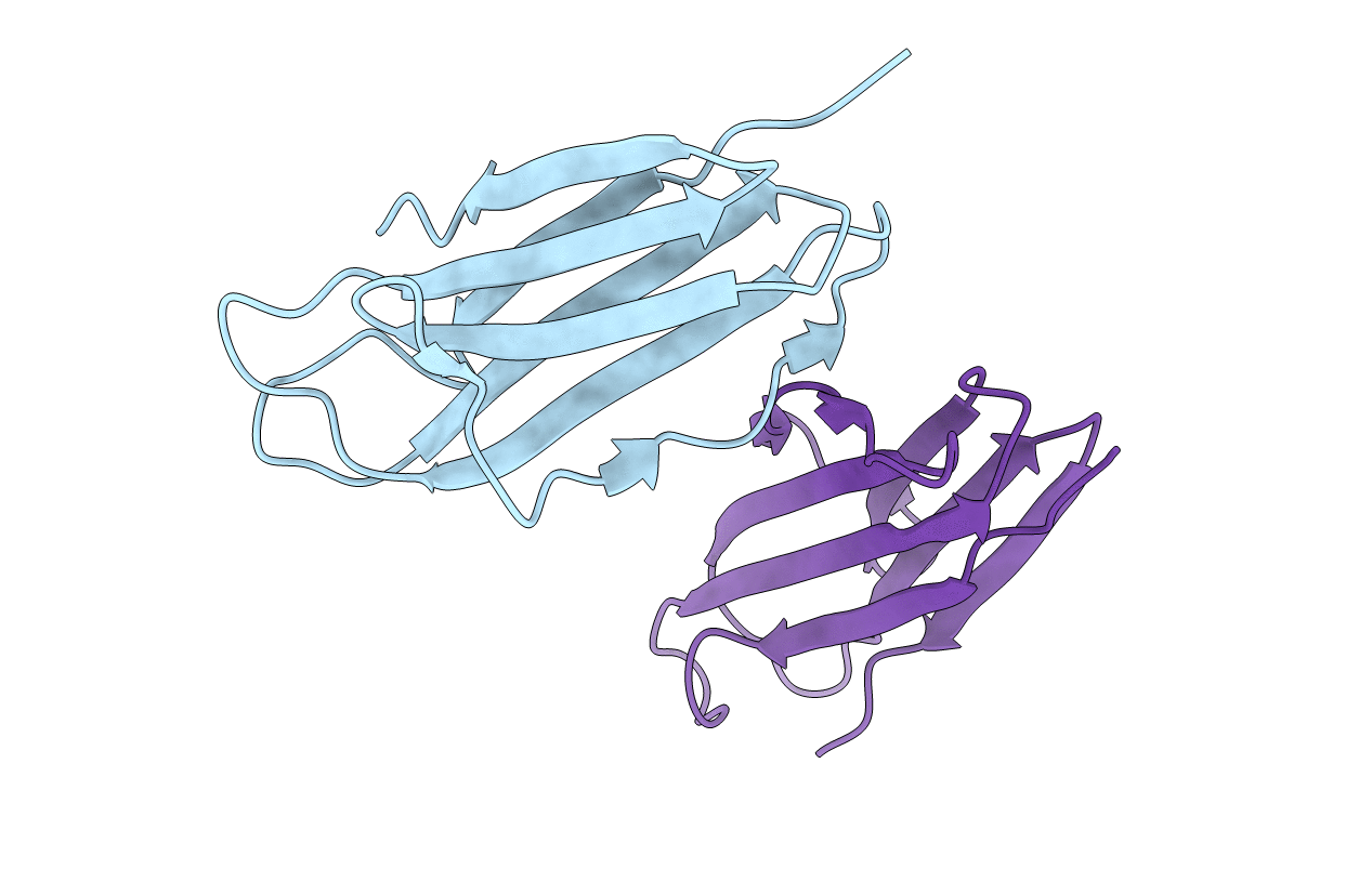
Structure of nurse shark beta-2-microglobulin
Biological Source:
Source Organism:
Host Organism:
Method Details:
Experimental Method:
Resolution:
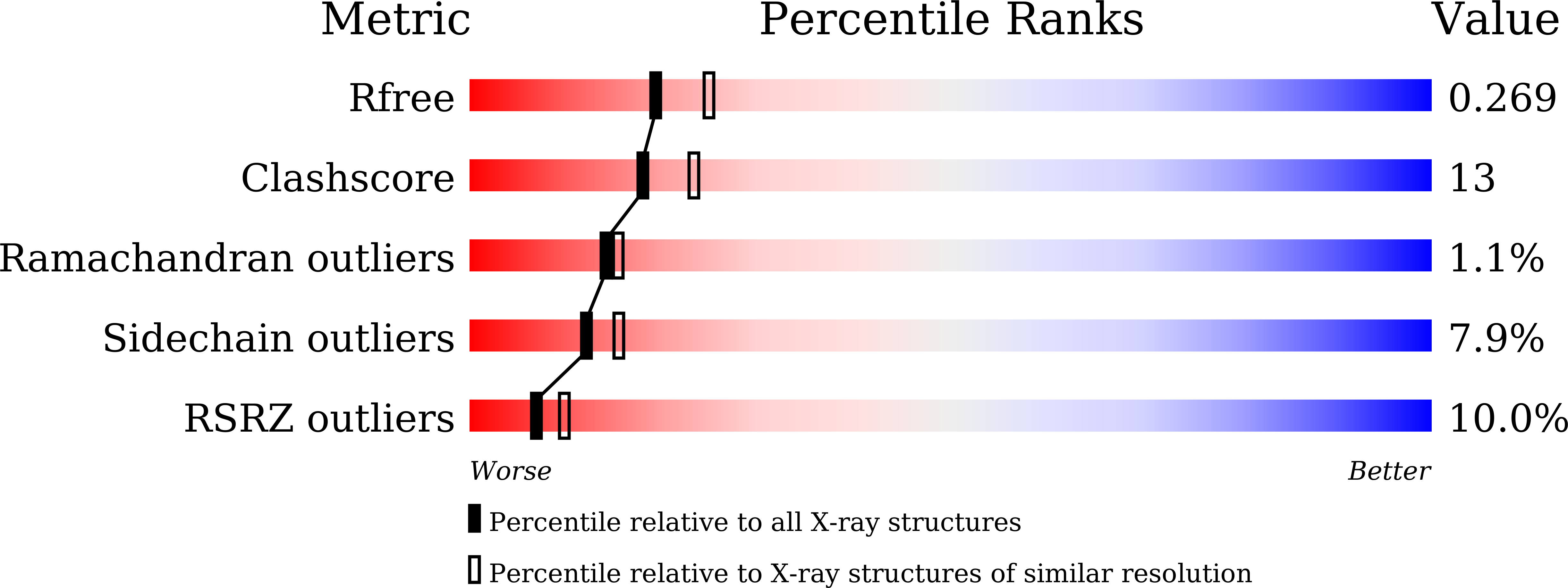
2.30 Å
R-Value Free:
0.26
R-Value Work:
0.23
Space Group:
P 32 2 1
Macromolecular Entities
Structures with similar UniProt ID
Protein Blast
Polymer Type:polypeptide(L)
Molecule:Beta-2-microglobulin
Gene (Uniprot):B2M
Chain IDs:A, B
Chain Length:96
Number of Molecules:2
Biological Source:Ginglymostoma cirratum
Primary Citation
The Structure of a Peptide-Loaded Shark MHC Class I Molecule Reveals Features of the Binding between beta 2 -Microglobulin and H Chain Conserved in Evolution.
J Immunol. ? ? ? (2021)
PMID: 34145057 DOI: 10.4049/jimmunol.2001165

Abstact

Cartilaginous fish are the most primitive extant species with MHC molecules. Using the nurse shark, the current study is, to the best of our knowledge, the first to present a peptide-loaded MHC class I (pMHC-I) structure for this class of animals. The overall structure was found to be similar between cartilaginous fish and bony animals, showing remarkable conservation of interactions between the three pMHC-I components H chain, β2-microglobulin (β2-m), and peptide ligand. In most previous studies, relatively little attention was given to the details of binding between the H chain and β2-m, and our study provides important new insights. A pronounced conserved feature involves the insertion of a large β2-m F56+W60 hydrophobic knob into a pleat of the β-sheet floor of the H chain α1α2 domain, with the knob being surrounded by conserved residues. Another conserved feature is a hydrogen bond between β2-m Y10 and a proline in the α3 domain of the H chain. By alanine substitution analysis, we found that the conserved β2-m residues Y10, D53, F56, and W60-each binding the H chain-are required for stable pMHC-I complex formation. For the β2-m residues Y10 and F56, such observations have not been reported before. The combined data indicate that for stable pMHC-I complex formation β2-m should not only bind the α1α2 domain but also the α3 domain. Knowing the conserved structural features of pMHC-I should be helpful for future elucidations of the mechanisms of pMHC-I complex formation and peptide editing.

Legend

Protein

Chemical

Disease

Primary Citation of related structures