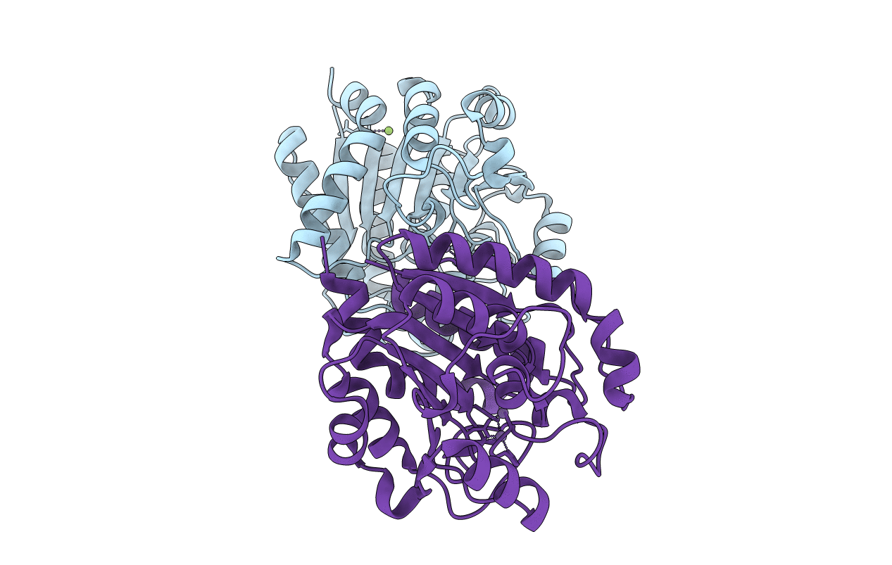
6LUH image
Entry Detail
PDB ID:
6LUH
Keywords:
Title:
High resolution structure of N(omega)-hydroxy-L-arginine hydrolase
Biological Source:
Source Organism:
PDB Version:
Deposition Date:
2020-01-28
Release Date:
2020-09-02
Method Details:
Experimental Method:
Resolution:
1.50 Å
R-Value Free:
0.23
R-Value Work:
0.19
R-Value Observed:
0.20
Space Group:
P 1
Macromolecular Entities
Polymer Type:polypeptide(L)
Description:N(omega)-hydroxy-L-arginine amidinohydrolase
Chain IDs:A, B
Chain Length:281
Number of Molecules:2
Biological Source:Streptomyces lavendulae
Primary Citation
Crystal structure of an Nomega-hydroxy-L-arginine hydrolase found in the D-cycloserine biosynthetic pathway.
Acta Crystallogr D Struct Biol 76 506 514 (2020)
PMID: 32496212 DOI: 10.1107/S2059798320004908

Abstact

DcsB, one of the enzymes encoded in the D-cycloserine (D-CS) biosynthetic gene cluster, displays a high sequence homology to arginase, which contains two manganese ions in the active site. However, DcsB hydrolyzes Nω-hydroxy-L-arginine, but not L-arginine, to supply hydroxyurea for the biosynthesis of D-CS. Here, the crystal structure of DcsB was determined at a resolution of 1.5 Å using anomalous scattering from the manganese ions. In the crystal structure, DscB generates an artificial dimer created by the open and closed forms. Gel-filtration analysis demonstrated that DcsB is a monomeric protein, unlike arginase, which forms a trimeric structure. The active center containing the binuclear manganese cluster differs between DcsB and arginase. In DcsB, one of the ligands of the MnA ion is a cysteine, while the corresponding residue in arginase is a histidine. In addition, DcsB has no counterpart to the histidine residue that acts as a general acid/base during the catalytic reaction of arginase. The present study demonstrates that DcsB has a unique active site that differs from that of arginase.

Legend

Protein

Chemical

Disease

Primary Citation of related structures