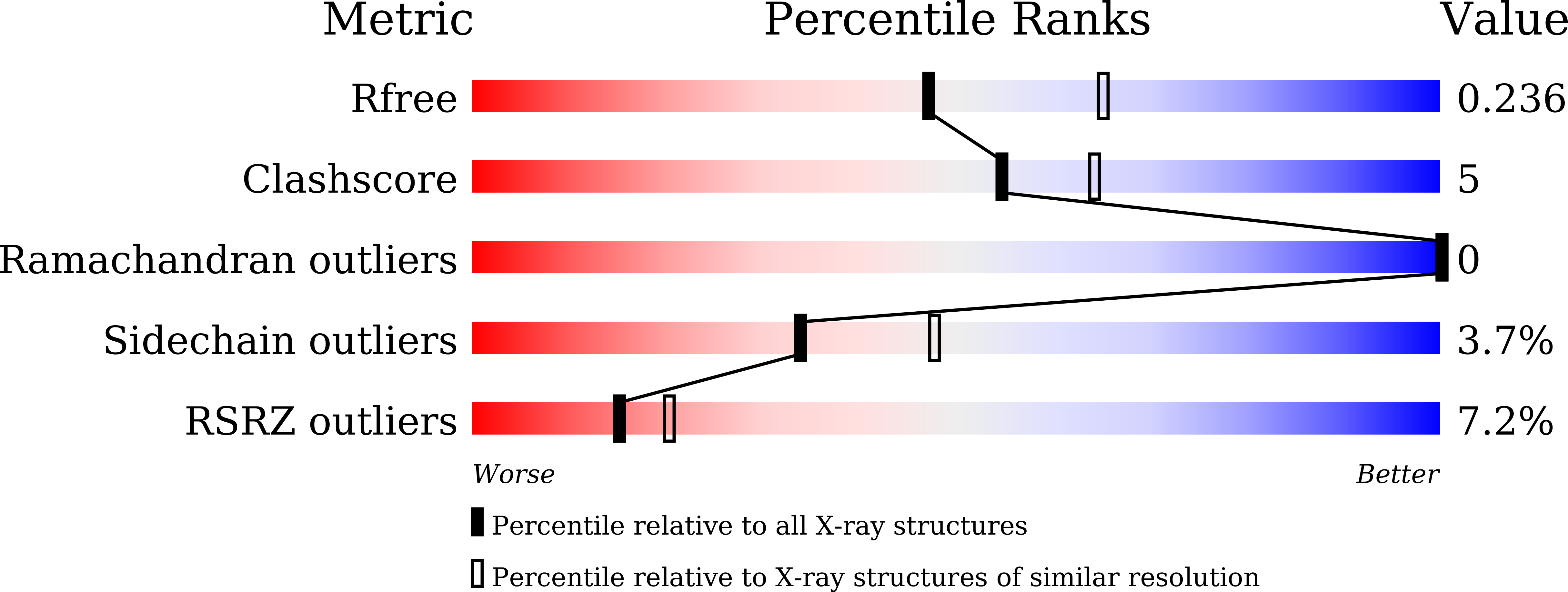
6LPG image
Deposition Date 2020-01-10
Release Date 2020-10-21
Last Version Date 2023-11-29
Entry Detail
PDB ID:
6LPG
Keywords:
Title:
human VASH1-SVBP complex
Biological Source:
Source Organism:
Homo sapiens (Taxon ID: 9606)
Method Details:
Experimental Method:
Resolution:
2.30 Å
R-Value Free:
0.23
R-Value Work:
0.18
R-Value Observed:
0.19
Space Group:
P 61 2 2
Macromolecular Entities
Structures with similar UniProt ID
Protein Blast
Polymer Type:polypeptide(L)
Molecule:Tubulinyl-Tyr carboxypeptidase 1
Gene (Uniprot):VASH1
Chain IDs:A
Chain Length:256
Number of Molecules:1
Biological Source:Homo sapiens
Structures with similar UniProt ID
Protein Blast
Polymer Type:polypeptide(L)
Molecule:Small vasohibin-binding protein
Gene (Uniprot):SVBP
Chain IDs:B
Chain Length:67
Number of Molecules:1
Biological Source:Homo sapiens
Ligand Molecules
Primary Citation
The crystal structure of the tetrameric human vasohibin-1-SVBP complex reveals a variable arm region within the structural core.
Acta Crystallogr D Struct Biol 76 993 1000 (2020)
PMID: 33021501 DOI: 10.1107/S2059798320011298

Abstact

Vasohibins regulate angiogenesis, tumor growth, metastasis and neuronal differentiation. They form a complex with small vasohibin-binding protein (SVBP) and show tubulin tyrosine carboxypeptidase activity. Recent crystal structure determinations of vasohibin-SVBP complexes have provided a molecular basis for complex formation, substrate binding and catalytic activity. However, the regulatory mechanism and dynamics of the complex remain elusive. Here, the crystal structure of the VASH1-SVBP complex and a molecular-dynamics simulation study are reported. The overall structure of the complex was similar to previously reported structures. Importantly, however, the structure revealed a domain-swapped heterotetramer that was formed between twofold symmetry-related molecules. This heterotetramerization was stabilized by the mutual exchange of ten conserved N-terminal residues from the VASH1 structural core, which was intramolecular in other structures. Interestingly, a comparison of this region with previously reported structures revealed that the patterns of hydrogen bonding and hydrophobic interactions vary. In the molecular-dynamics simulations, differences were found between the heterotetramer and heterodimer, where the fluctuation of the N-terminal region in the heterotetramer was suppressed. Thus, heterotetramer formation and flexibility of the N-terminal region may be important for enzyme activity and regulation.

Legend

Protein

Chemical

Disease

Primary Citation of related structures