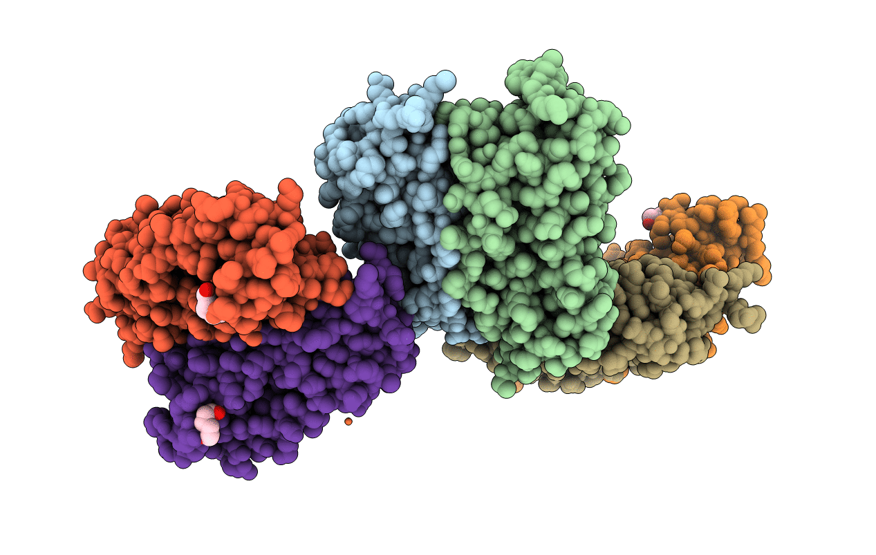
6LP5 image
Deposition Date 2020-01-08
Release Date 2020-04-08
Last Version Date 2023-11-29
Entry Detail
PDB ID:
6LP5
Keywords:
Title:
Structure of Sinonovacula constricta ferritin
Biological Source:
Source Organism:
Method Details:
Experimental Method:
Resolution:
1.98 Å
R-Value Free:
0.20
R-Value Work:
0.16
R-Value Observed:
0.16
Space Group:
I 4
Macromolecular Entities
Polymer Type:polypeptide(L)
Molecule:Ferritin
Chain IDs:A, B, C, D, E, F
Chain Length:171
Number of Molecules:6
Biological Source:Sinonovacula constricta
Primary Citation
Crystallographic characterization of ferritin from Sinonovacula constricta.
Biochem.Biophys.Res.Commun. 524 217 223 (2020)
PMID: 31983429 DOI: 10.1016/j.bbrc.2020.01.069

Abstact

Ferritins are ubiquitous iron-binding proteins that are mainly related to iron storage, detoxification and innate immunity. Here, we present the crystal structure of a marine invertebrate ferritin from Sinonovacula constricta at a resolution of 1.98 Å. The S. constricta ferritin (ScFer) possessed some structural similarities with vertebrate ferritins, and they shared a well-conserved architecture composed of five α-helical bundles that assembled into a cage-like structure with 24-subunits. The structure of ScFer also showed iron binding sites in the 3-fold channel, ferroxidase center, and putative nucleation sites. Further, electrostatic potential calculations suggested that the electrostatic gradient of the 3-fold channel could provide a guidance mechanism for iron entering the ferritin cavity.

Legend

Protein

Chemical

Disease

Primary Citation of related structures