Deposition Date
2020-01-07
Release Date
2020-09-02
Last Version Date
2023-11-29
Entry Detail
PDB ID:
6LOS
Keywords:
Title:
Crystal structure of mouse PEDF in complex with heterotrimeric collagen model peptide.
Biological Source:
Source Organism(s):
Mus musculus (Taxon ID: 10090)
Homo sapiens (Taxon ID: 9606)
Homo sapiens (Taxon ID: 9606)
Expression System(s):
Method Details:
Experimental Method:
Resolution:
2.48 Å
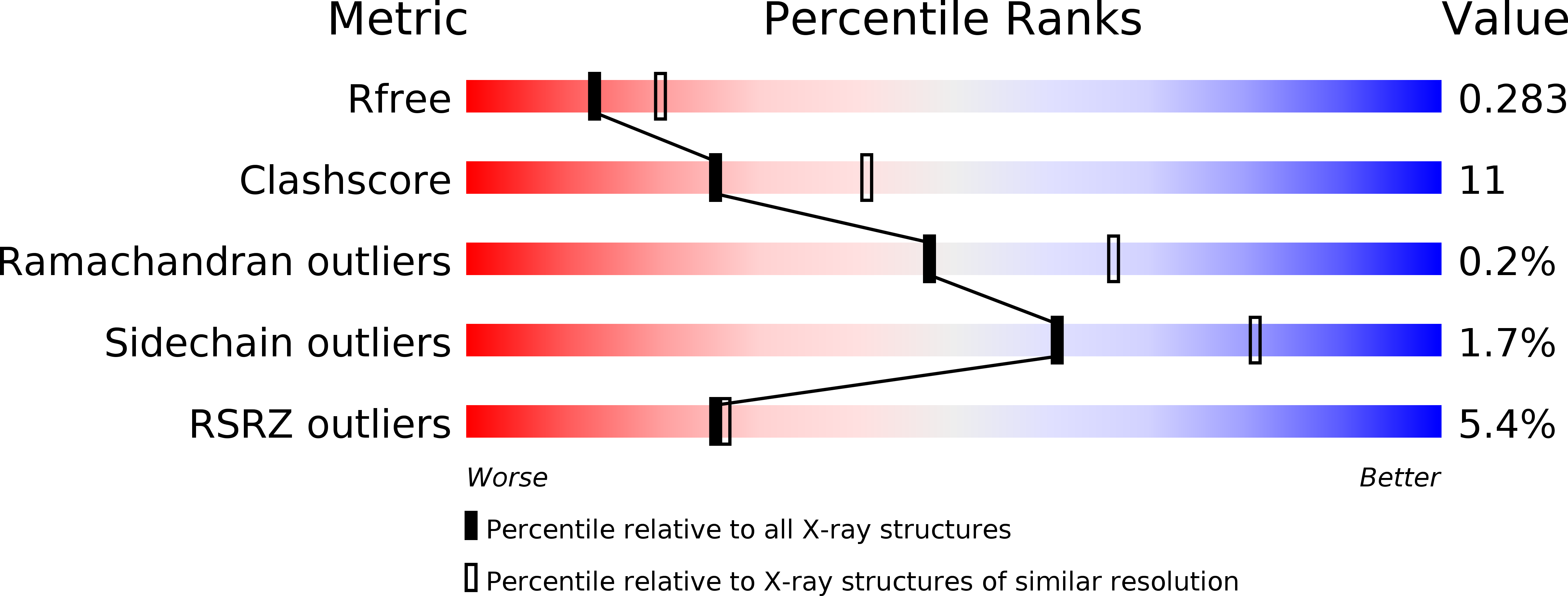
R-Value Free:
0.28
R-Value Work:
0.22
R-Value Observed:
0.22
Space Group:
C 2 2 21