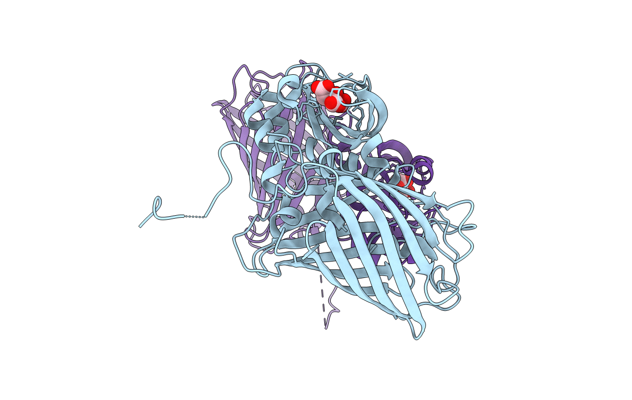
6LNP image
Deposition Date 2019-12-31
Release Date 2020-05-06
Last Version Date 2024-10-09
Entry Detail
PDB ID:
6LNP
Title:
Crystal structure of citrate Biosensor
Biological Source:
Source Organism:
Host Organism:
Method Details:
Experimental Method:
Resolution:
2.99 Å
R-Value Free:
0.28
R-Value Work:
0.22
R-Value Observed:
0.22
Space Group:
P 41
Macromolecular Entities
Polymer Type:polypeptide(L)
Molecule:Fusion protein of Green fluorescent protein and Sensor histidine kinase CitA
Gene (Uniprot):citA
Chain IDs:A (auth: B), B (auth: D)
Chain Length:405
Number of Molecules:2
Biological Source:Aequorea victoria, Klebsiella pneumoniae
Ligand Molecules
Primary Citation
High-Performance Intensiometric Direct- and Inverse-Response Genetically Encoded Biosensors for Citrate.
Acs Cent.Sci. 6 1441 1450 (2020)
PMID: 32875085 DOI: 10.1021/acscentsci.0c00518

Abstact

Motivated by the growing recognition of citrate as a central metabolite in a variety of biological processes associated with healthy and diseased cellular states, we have developed a series of high-performance genetically encoded citrate biosensors suitable for imaging of citrate concentrations in mammalian cells. The design of these biosensors was guided by structural studies of the citrate-responsive sensor histidine kinase and took advantage of the same conformational changes proposed to propagate from the binding domain to the catalytic domain. Following extensive engineering based on a combination of structure guided mutagenesis and directed evolution, we produced an inverse-response biosensor (ΔF/F min ≈ 18) designated Citroff1 and a direct-response biosensor (ΔF/F min ≈ 9) designated Citron1. We report the X-ray crystal structure of Citron1 and demonstrate the utility of both biosensors for qualitative and quantitative imaging of steady-state and pharmacologically perturbed citrate concentrations in live cells.

Legend

Protein

Chemical

Disease

Primary Citation of related structures