6LJE image
Deposition Date 2019-12-14
Release Date 2020-01-01
Last Version Date 2023-11-22
Entry Detail
PDB ID:
6LJE
Title:
Crystal structure of gelsolin G3 domain (calcium and magnesium condition)
Biological Source:
Source Organism:
Homo sapiens (Taxon ID: 9606)
Host Organism:
Method Details:
Experimental Method:
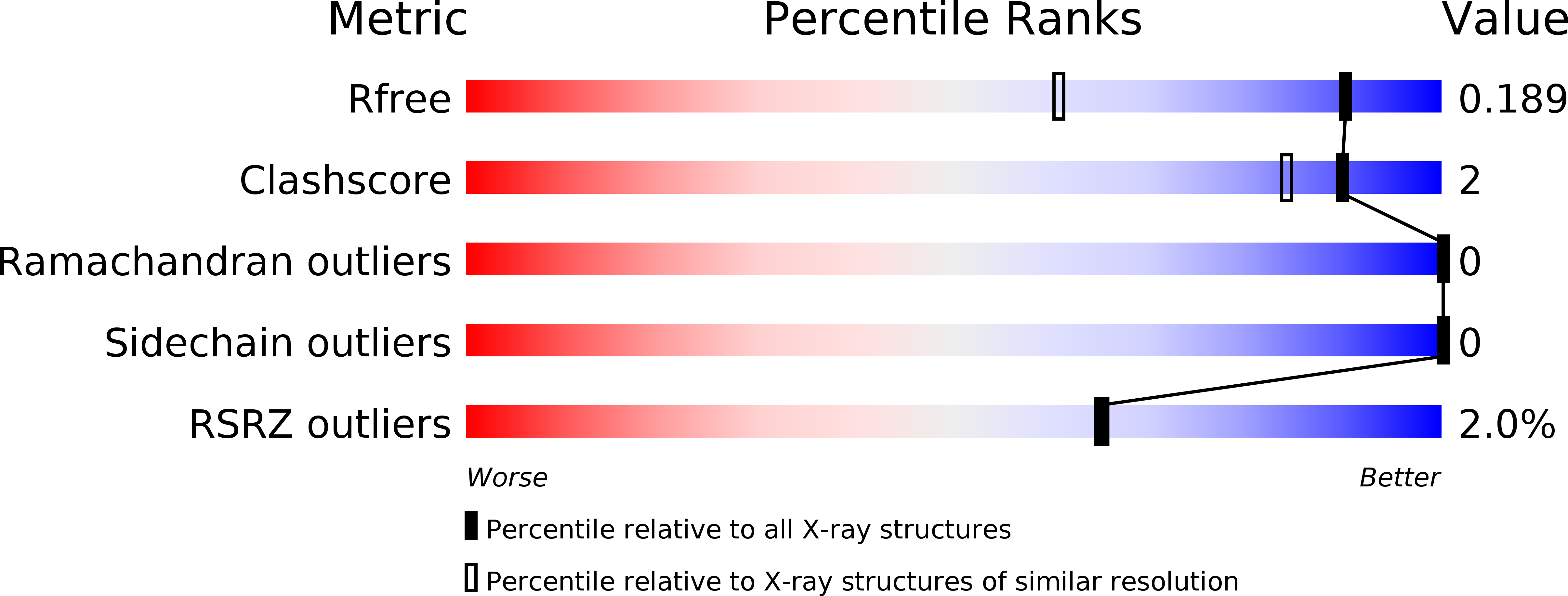
Resolution:
1.40 Å
R-Value Free:
0.18
R-Value Work:
0.18
R-Value Observed:
0.18
Space Group:
C 1 2 1
Macromolecular Entities
Polymer Type:polypeptide(L)
Molecule:Gelsolin
Gene (Uniprot):GSN
Chain IDs:A, B
Chain Length:101
Number of Molecules:2
Biological Source:Homo sapiens
Primary Citation
Novel inter-domain Ca2+-binding site in the gelsolin superfamily protein fragmin.
J.Muscle Res.Cell.Motil. 41 153 162 (2020)
PMID: 31863323 DOI: 10.1007/s10974-019-09571-5

Abstact

Gelsolin superfamily proteins, consisting of multiple domains (usually six), sever actin filaments and cap the barbed ends in a Ca2+-dependent manner. Two types of evolutionally conserved Ca2+-binding sites have been identified in this family; type-1 (between gelsolin and actin) and type-2 (within the gelsolin domain). Fragmin, a member in the slime mold Physarum polycephalum, consists of three domains (F1-F3) that are highly similar to the N-terminal half of mammalian gelsolin (G1-G3). Despite their similarities, the two proteins exhibit a significant difference in the Ca2+ dependency; F1-F3 absolutely requires Ca2+ for the filament severing whereas G1-G3 does not. In this study, we examined the strong dependency of fragmin on Ca2+ using biochemical and structural approaches. Our co-sedimentation assay demonstrated that Ca2+ significantly enhanced the binding of F2-F3 to actin. We determined the crystal structure of F2-F3 in the presence of Ca2+. F2-F3 binds a total of three calcium ions; while two are located in type-2 sites within F2 or F3, the remaining one resides between the F2 long helix and the F3 short helix. The inter-domain Ca2+-coordination appears to stabilize F2-F3 in a closely packed configuration. Notably, the F3 long helix exhibits a bent conformation which is different from the straight G3 long helix in the presence of Ca2+. Our results provide the first structural evidence for the existence of an unconventional Ca2+-binding site in the gelsolin superfamily proteins.

Legend

Protein

Chemical

Disease

Primary Citation of related structures