6LIO image
Deposition Date 2019-12-12
Release Date 2020-09-30
Last Version Date 2023-11-22
Entry Detail
PDB ID:
6LIO
Keywords:
Title:
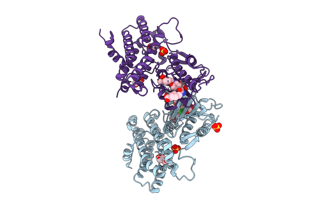
Crystal structure of human PDK2 complexed with GM67520
Biological Source:
Source Organism:
Homo sapiens (Taxon ID: 9606)
Method Details:
Experimental Method:
Resolution:
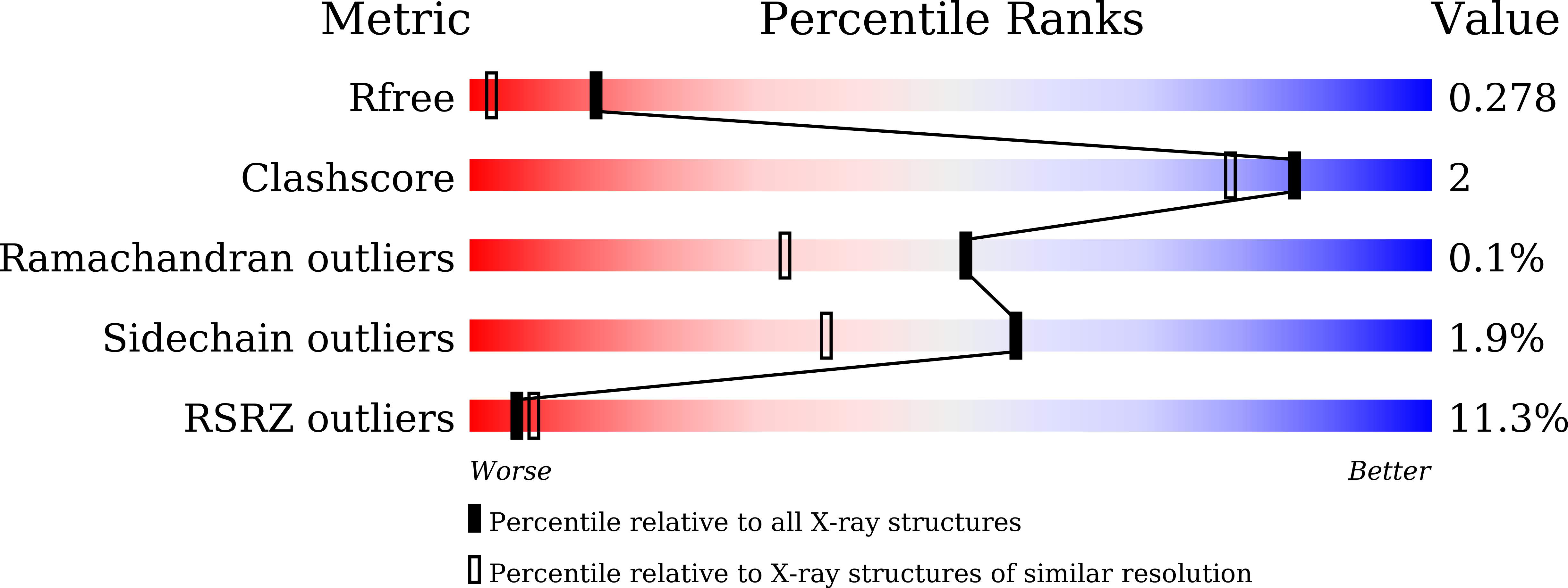
1.76 Å
R-Value Free:
0.26
R-Value Work:
0.23
R-Value Observed:
0.23
Space Group:
P 21 21 21
Macromolecular Entities
Polymer Type:polypeptide(L)
Molecule:[Pyruvate dehydrogenase (acetyl-transferring)] kinase isozyme 2, mitochondrial
Gene (Uniprot):PDK2
Chain IDs:A, B
Chain Length:411
Number of Molecules:2
Biological Source:Homo sapiens
Primary Citation
Structural basis for the inhibition of PDK2 by novel ATP- and lipoyl-binding site targeting compounds.
Biochem.Biophys.Res.Commun. 527 778 784 (2020)
PMID: 32444142 DOI: 10.1016/j.bbrc.2020.04.102

Abstact

Pyruvate dehydrogenase kinase (PDK) controls the activity of pyruvate decarboxylase complex (PDC) by phosphorylating key serine residues on the E1 subunit, which leads to a decreased oxidative phosphorylation in mitochondria. Inhibition of PDK activity by natural/synthetic compounds has been shown to reverse the Warburg effect, a characteristic metabolism in cancer cells. PDK-PDC axis also has been associated with diabetes and heart disease. Therefore, regulation of PDK activity has been considered as a promising strategy to treat related diseases. Here we present the X-ray crystal structure of PDK2 complexed with a recently identified PDK4 inhibitor, compound 8c, which has been predicted to bind at the lipoyl-binding site and interrupt intermolecular interactions with the E2-E3bp subunits of PDC. The co-crystal structure confirmed the specific binding location of compound 8c and revealed the remote conformational change in the ATP-binding pocket. In addition, two novel 4,5-diarylisoxazole derivatives, GM10030 and GM67520, were synthesized and used for structural studies, which target the ATP-binding site of PDK2. These compounds bind to PDK2 with a sub-100nM affinity as determined by isothermal titration calorimetry experiments. Notably, the crystal structure of the PDK2-GM10030 complex displays unprecedented asymmetric conformation of human PDK2 dimer, especially in the ATP-lids and C-terminal tails.

Legend

Protein

Chemical

Disease

Primary Citation of related structures