Deposition Date
2019-11-12
Release Date
2020-06-10
Last Version Date
2023-11-22
Entry Detail
PDB ID:
6LAA
Keywords:
Title:
Crystal structure of full-length CYP116B46 from Tepidiphilus thermophilus
Biological Source:
Source Organism(s):
Tepidiphilus thermophilus (Taxon ID: 876478)
Expression System(s):
Method Details:
Experimental Method:
Resolution:
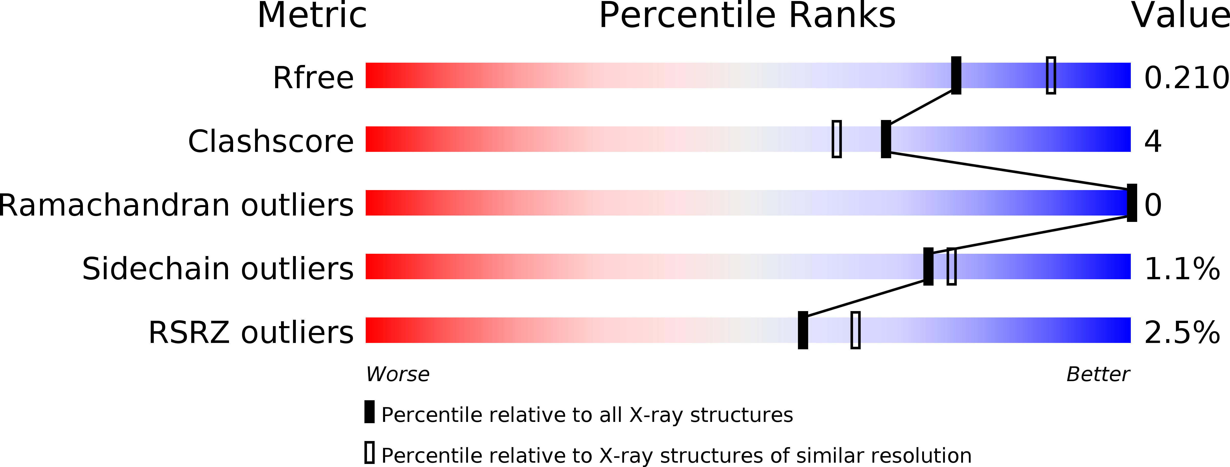
2.13 Å
R-Value Free:
0.21
R-Value Work:
0.16
R-Value Observed:
0.16
Space Group:
P 43 21 2