6L9S image
Deposition Date 2019-11-10
Release Date 2020-01-29
Last Version Date 2023-11-22
Entry Detail
PDB ID:
6L9S
Title:
Crystal structure of Na-dithionite reduced auracyanin from photosynthetic bacterium Roseiflexus castenholzii
Biological Source:
Method Details:
Experimental Method:
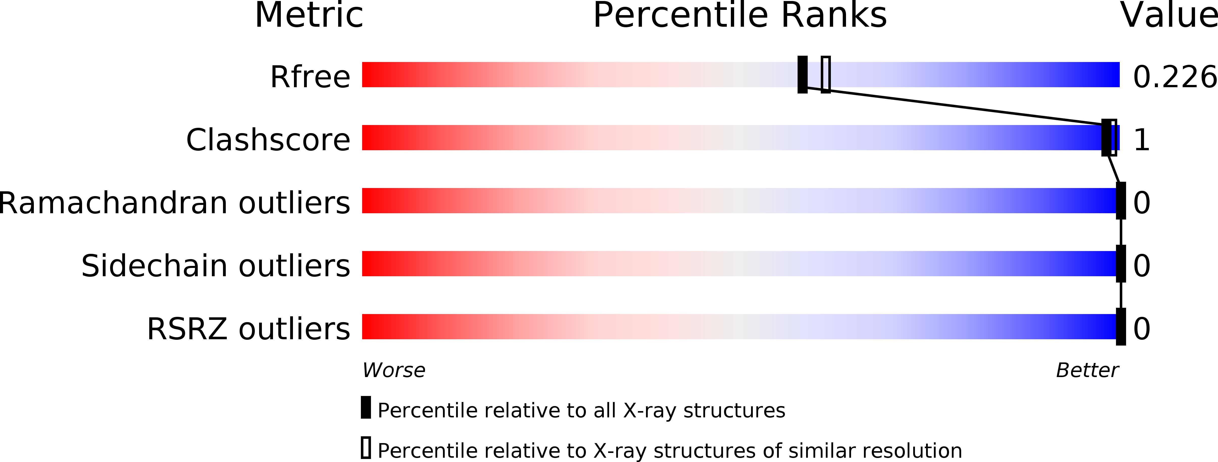
Resolution:
2.00 Å
R-Value Free:
0.21
R-Value Work:
0.17
R-Value Observed:
0.17
Space Group:
P 2 21 21
Macromolecular Entities
Structures with similar UniProt ID
Protein Blast
Polymer Type:polypeptide(L)
Molecule:Blue (Type 1) copper domain protein
Gene (Uniprot):Rcas_3112
Chain IDs:A
Chain Length:129
Number of Molecules:1
Biological Source:Roseiflexus castenholzii (strain DSM 13941 / HLO8)
Ligand Molecules
Primary Citation
Structural basis underlying the electron transfer features of a blue copper protein auracyanin from the photosynthetic bacterium Roseiflexus castenholzii.
Photosyn. Res. 143 301 314 (2020)
PMID: 31933173 DOI: 10.1007/s11120-020-00709-y

Abstact

Auracyanin (Ac) is a blue copper protein that mediates the electron transfer between Alternative Complex III (ACIII) and downstream electron acceptors in both fort chains of filamentous anoxygenic phototrophs. Here, we extracted and purified the air-oxidized RfxAc from the photoheterotrophically grown Roseiflexus castenholzii, and we illustrated the structural basis underlying its electron transferring features. Spectroscopic and enzymatic analyses demonstrated the reduction of air-oxidized RfxAc by the ACIII upon oxidation of menaquinol-4 and menaquinol-7. Crystal structures of the air-oxidized and Na-dithionite-reduced RfxAc at 2.2 and 2.0 Å resolutions, respectively, showed that the copper ions are coordinated by His77, His146, Cys141, and Met151 in minor different geometries. The Cu1-Sδ bond length increase of Met151, and the electron density Fourier differences at Cu1 and His77 demonstrated their essential roles in the dithionite-induced reduction. Structural comparisons further revealed that the RfxAc contains a Chloroflexus aurantiacus Ac-A-like copper binding pocket and a hydrophobic patch surrounding the exposed edge of His146 imidazole, as well as an Ac-B-like Ser- and Thr-rich polar patch located at a different site on the surface. These spectroscopic and structural features allow RfxAc to mediate electron transfers between the ACIII and redox partners different from those of Ac-A and Ac-B. These results provide a structural basis for further investigating the electron transfer and energy transformation mechanism of bacterial photosynthesis, and the diversity and evolution of electron transport chains.

Legend

Protein

Chemical

Disease

Primary Citation of related structures