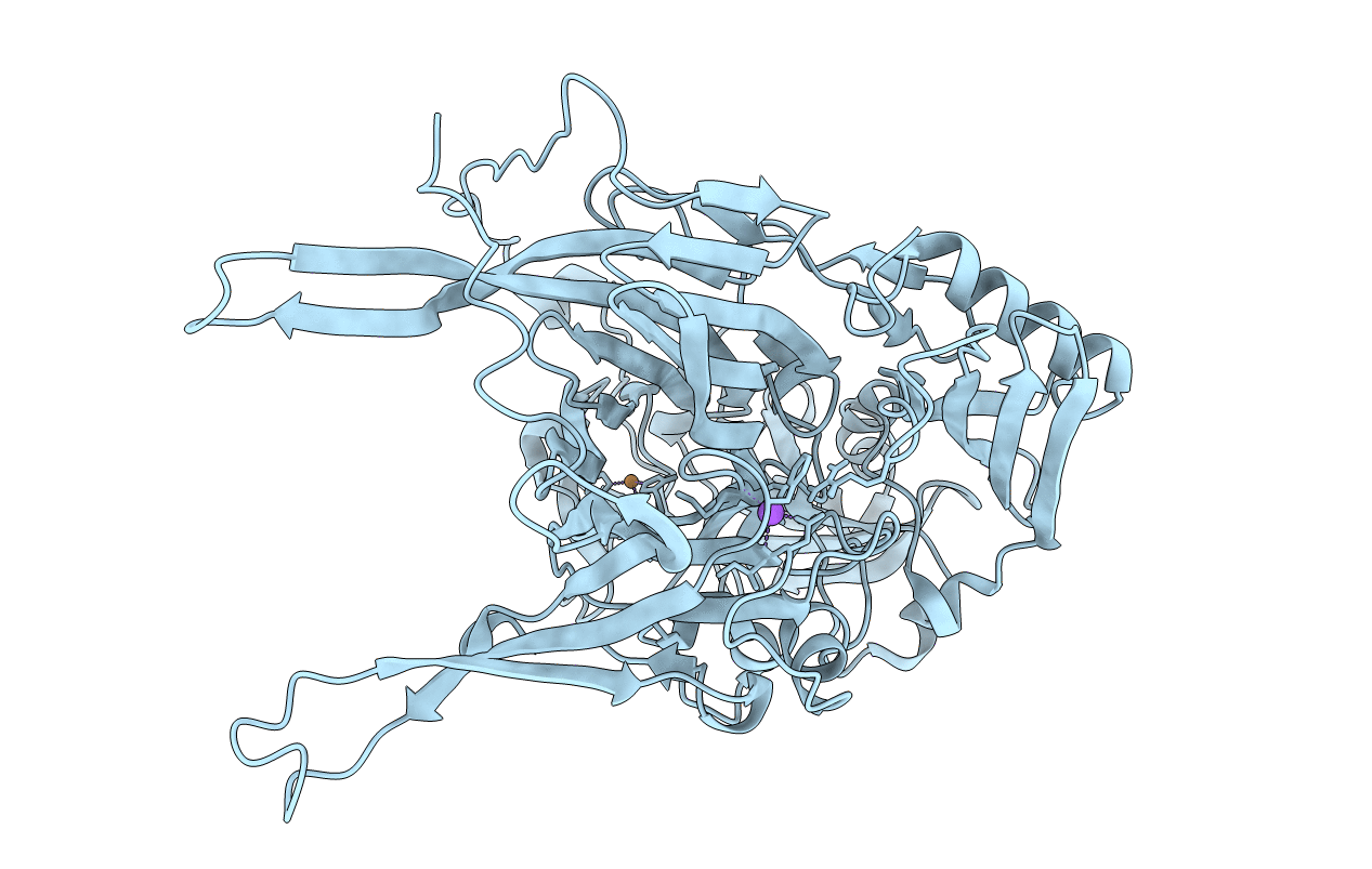
6L9C image
Deposition Date 2019-11-08
Release Date 2020-04-29
Last Version Date 2023-11-22
Entry Detail
PDB ID:
6L9C
Keywords:
Title:
Neutron structure of copper amine oxidase from Arthrobacter glibiformis at pD 7.4
Biological Source:
Source Organism:
Host Organism:
Method Details:
R-Value Free:
['0.18
R-Value Work:
['0.16
R-Value Observed:
['0.16
Space Group:
C 1 2 1
Macromolecular Entities
Structures with similar UniProt ID
Protein Blast
Polymer Type:polypeptide(L)
Molecule:Phenylethylamine oxidase
Chain IDs:A (auth: X)
Chain Length:621
Number of Molecules:1
Biological Source:Arthrobacter globiformis
Modified Residue
Compound ID Chain ID Parent Comp ID Details 2D Image
ASA A ASP modified residue
E9C A TYR modified residue
Primary Citation
Neutron crystallography of copper amine oxidase reveals keto/enolate interconversion of the quinone cofactor and unusual proton sharing.
Proc.Natl.Acad.Sci.USA 117 10818 10824 (2020)
PMID: 32371483 DOI: 10.1073/pnas.1922538117

Abstact

Recent advances in neutron crystallographic studies have provided structural bases for quantum behaviors of protons observed in enzymatic reactions. Thus, we resolved the neutron crystal structure of a bacterial copper (Cu) amine oxidase (CAO), which contains a prosthetic Cu ion and a protein-derived redox cofactor, topa quinone (TPQ). We solved hitherto unknown structures of the active site, including a keto/enolate equilibrium of the cofactor with a nonplanar quinone ring, unusual proton sharing between the cofactor and the catalytic base, and metal-induced deprotonation of a histidine residue that coordinates to the Cu. Our findings show a refined active-site structure that gives detailed information on the protonation state of dissociable groups, such as the quinone cofactor, which are critical for catalytic reactions.

Legend

Protein

Chemical

Disease

Primary Citation of related structures