Deposition Date
2019-11-08
Release Date
2020-03-18
Last Version Date
2023-11-22
Entry Detail
PDB ID:
6L93
Keywords:
Title:
X-ray structure of the ligand-free human TRPV1 ankyrin repeat domain
Biological Source:
Source Organism(s):
Homo sapiens (Taxon ID: 9606)
Expression System(s):
Method Details:
Experimental Method:
Resolution:
4.47 Å
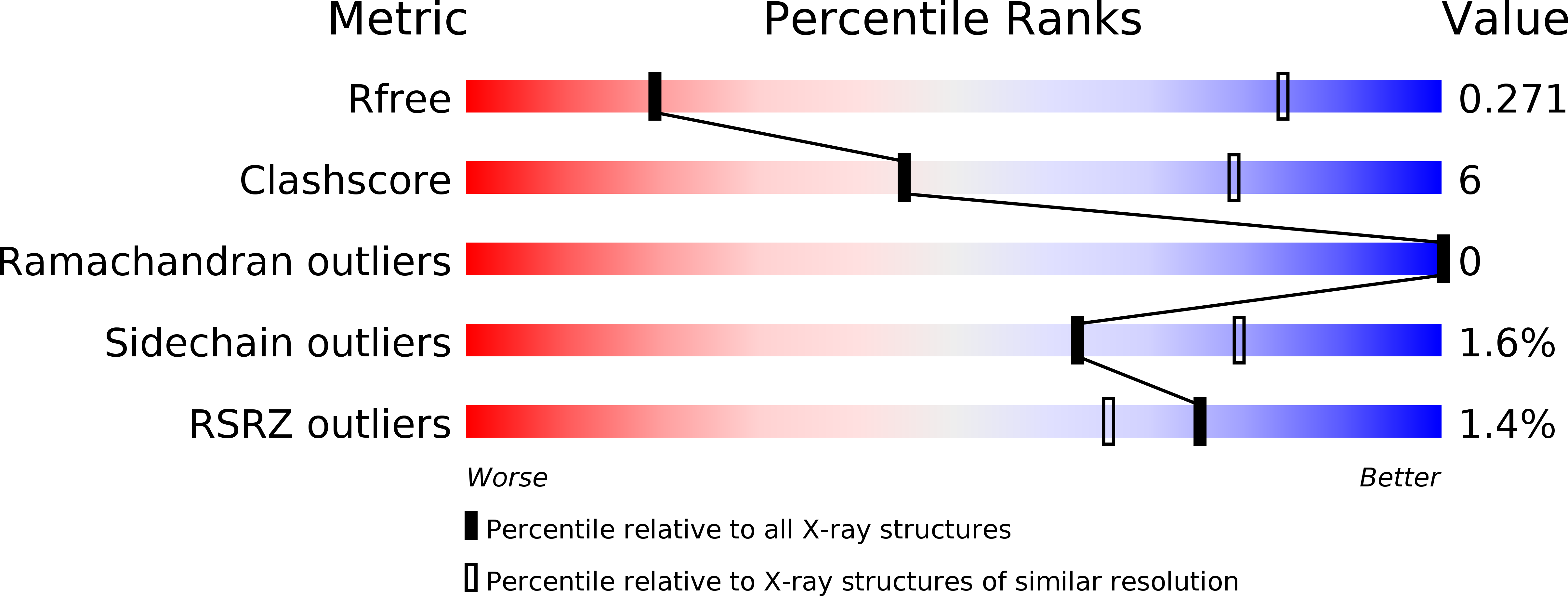
R-Value Free:
0.26
R-Value Work:
0.22
R-Value Observed:
0.22
Space Group:
C 1 2 1