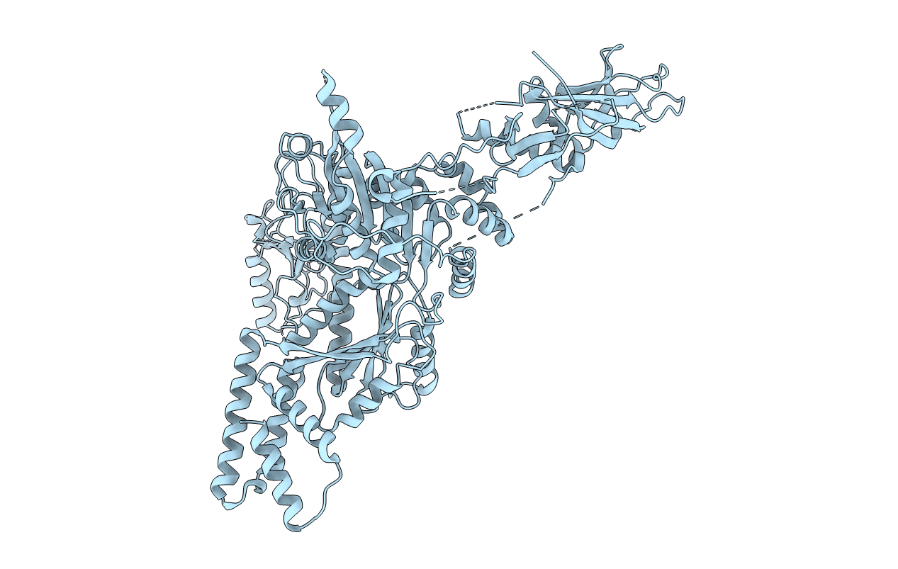
6L8N image
Deposition Date 2019-11-06
Release Date 2020-11-11
Last Version Date 2024-04-03
Entry Detail
PDB ID:
6L8N
Title:
Crystal structure of the K. lactis Rad5
Biological Source:
Source Organism:
Host Organism:
Method Details:
Experimental Method:
Resolution:
3.60 Å
R-Value Free:
0.23
R-Value Work:
0.20
R-Value Observed:
0.20
Space Group:
P 62 2 2
Macromolecular Entities
Structures with similar UniProt ID
Protein Blast
Polymer Type:polypeptide(L)
Molecule:DNA repair protein RAD5
Gene (Uniprot):RAD5
Chain IDs:A
Chain Length:952
Number of Molecules:1
Biological Source:Kluyveromyces lactis NRRL Y-1140
Ligand Molecules
Primary Citation
Structural basis for the multi-activity factor Rad5 in replication stress tolerance.
Nat Commun 12 321 321 (2021)
PMID: 33436623 DOI: 10.1038/s41467-020-20538-w

Abstact

The yeast protein Rad5 and its orthologs in other eukaryotes promote replication stress tolerance and cell survival using their multiple activities, including ubiquitin ligase, replication fork remodeling and DNA lesion targeting activities. Here, we present the crystal structure of a nearly full-length Rad5 protein. The structure shows three distinct, but well-connected, domains required for Rad5's activities. The spatial arrangement of these domains suggest that different domains can have autonomous activities but also undergo intrinsic coordination. Moreover, our structural, biochemical and cellular studies demonstrate that Rad5's HIRAN domain mediates interactions with the DNA metabolism maestro factor PCNA and contributes to its poly-ubiquitination, binds to DNA and contributes to the Rad5-catalyzed replication fork regression, defining a new type of HIRAN domains with multiple activities. Our work provides a framework to understand how Rad5 integrates its various activities in replication stress tolerance.

Legend

Protein

Chemical

Disease

Primary Citation of related structures