
Deposition Date
2019-10-07
Release Date
2020-05-13
Last Version Date
2024-03-27
Entry Detail
PDB ID:
6L2W
Keywords:
Title:
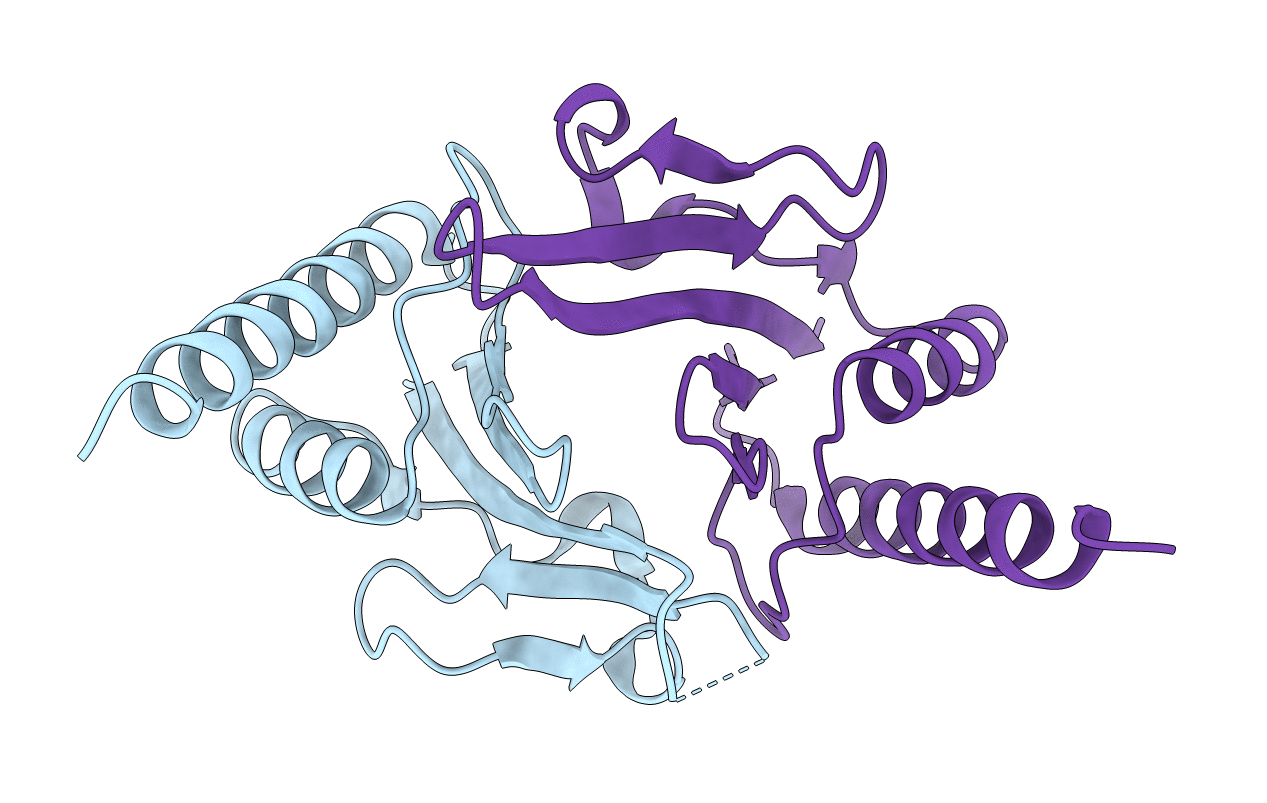
Crystal structure of a novel fold protein Gp72 from the freshwater cyanophage Mic1
Biological Source:
Source Organism(s):
Microcystis phage Mic1 (Taxon ID: 2587456)
Expression System(s):
Method Details:
Experimental Method:
Resolution:
2.29 Å
R-Value Free:
0.23
R-Value Work:
0.19
R-Value Observed:
0.20
Space Group:
C 1 2 1
