Deposition Date
2019-10-02
Release Date
2020-04-01
Last Version Date
2024-11-06
Entry Detail
PDB ID:
6L27
Keywords:
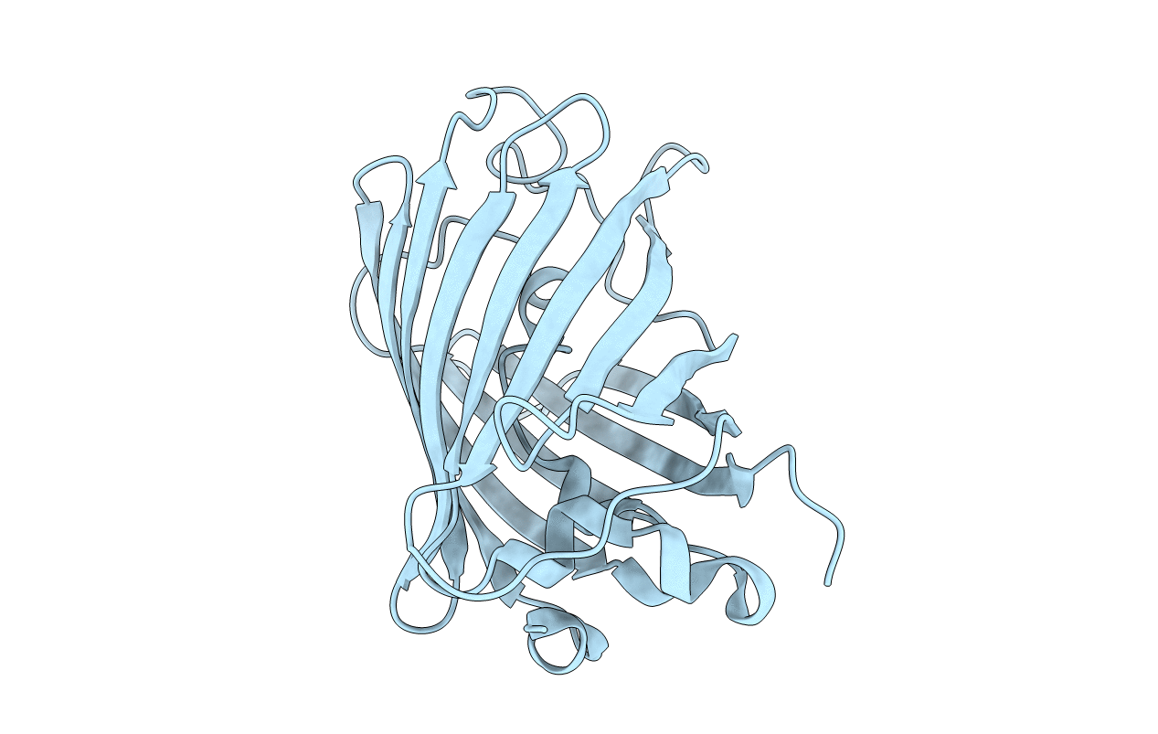
Title:
X-ray crystal structure of the mutant green fluorescent protein
Biological Source:
Source Organism(s):
Aequorea victoria (Taxon ID: 6100)
Expression System(s):
Method Details:
Experimental Method:
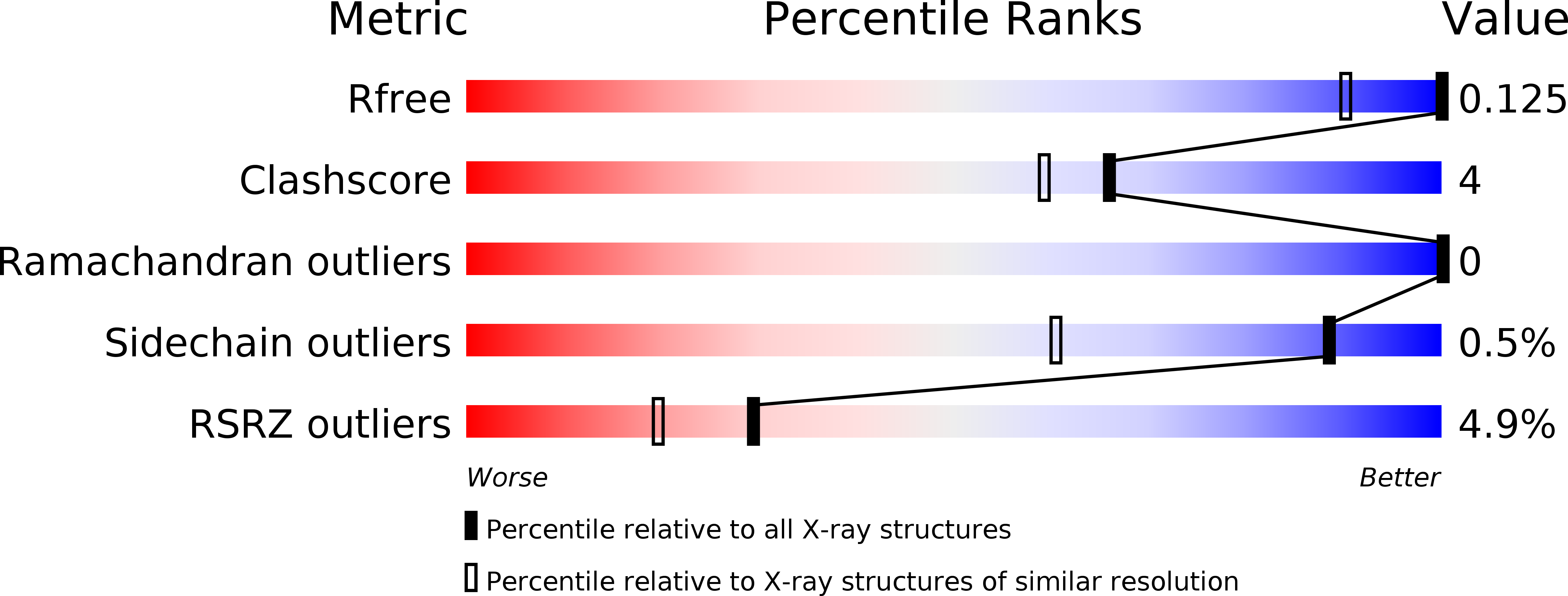
Resolution:
0.77 Å
R-Value Free:
0.12
R-Value Work:
0.11
R-Value Observed:
0.11
Space Group:
P 21 21 21