Deposition Date
2019-09-26
Release Date
2020-12-02
Last Version Date
2023-11-22
Entry Detail
PDB ID:
6L0K
Keywords:
Title:
Crystal structure of dihydroorotase in complex with malate at pH9 from Saccharomyces cerevisiae
Biological Source:
Source Organism(s):
Saccharomyces cerevisiae S288C (Taxon ID: 559292)
Expression System(s):
Method Details:
Experimental Method:
Resolution:
3.30 Å
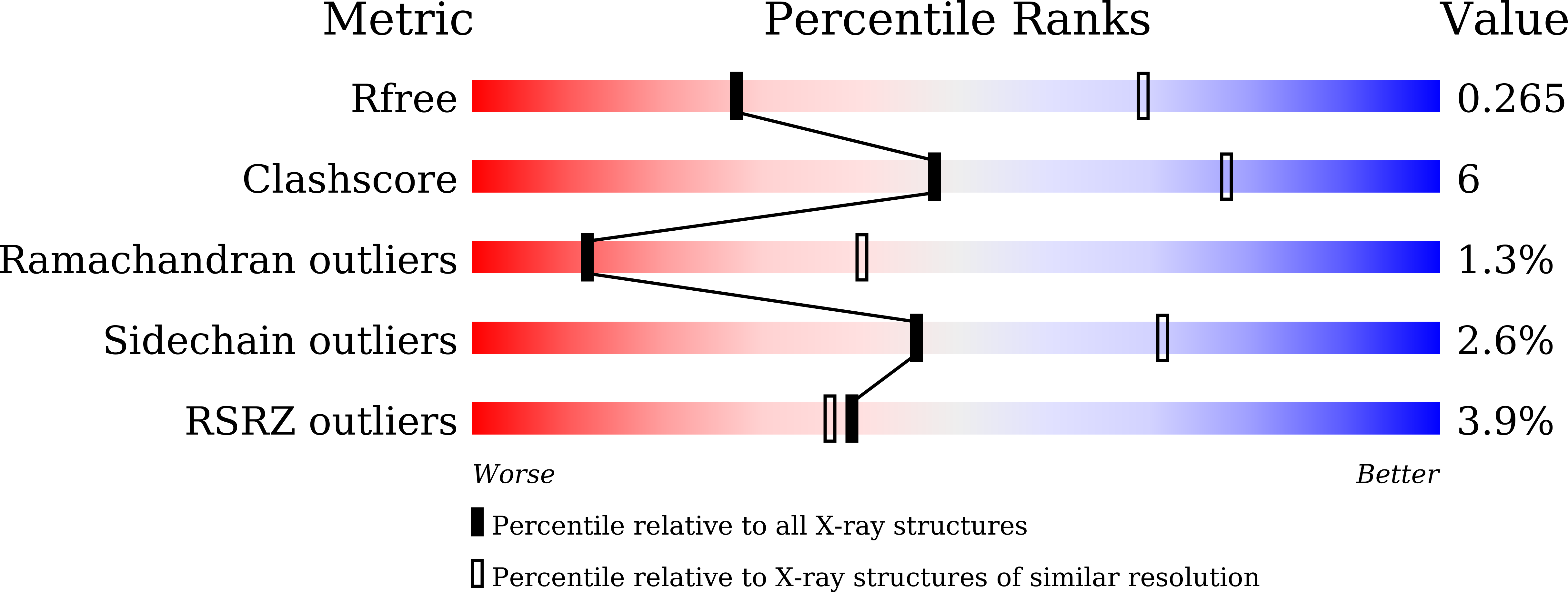
R-Value Free:
0.26
R-Value Work:
0.21
R-Value Observed:
0.21
Space Group:
P 1 21 1