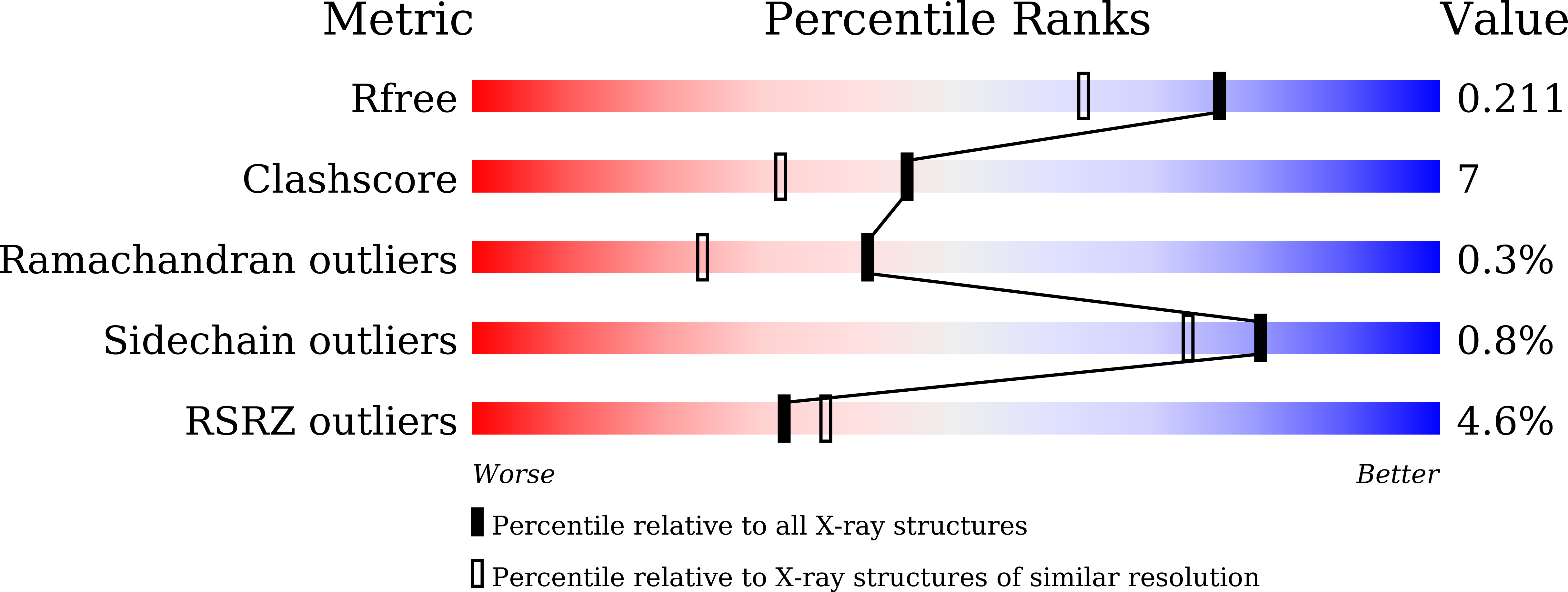
6KZQ image
Entry Detail
PDB ID:
6KZQ
Keywords:
Title:
structure of PTP-MEG2 and NSF-pY83 peptide complex
Biological Source:
Source Organism:
Host Organism:
PDB Version:
Deposition Date:
2019-09-25
Release Date:
2020-09-30
Method Details:
Experimental Method:
Resolution:
1.70 Å
R-Value Free:
0.20
R-Value Work:
0.18
R-Value Observed:
0.18
Space Group:
P 21 21 2
Macromolecular Entities
Polymer Type:polypeptide(L)
Description:Tyrosine-protein phosphatase non-receptor type 9
Chain IDs:A
Chain Length:307
Number of Molecules:1
Biological Source:Homo sapiens
Polymer Type:polypeptide(L)
Description:NSF-pY83 peptide
Chain IDs:B (auth: F)
Chain Length:9
Number of Molecules:1
Biological Source:Homo sapiens
Primary Citation
PTP-MEG2 regulates quantal size and fusion pore opening through two distinct structural bases and substrates.
Embo Rep. 22 e52141 e52141 (2021)
PMID: 33764618 DOI: 10.15252/embr.202052141

Abstact

Tyrosine phosphorylation of secretion machinery proteins is a crucial regulatory mechanism for exocytosis. However, the participation of protein tyrosine phosphatases (PTPs) in different exocytosis stages has not been defined. Here we demonstrate that PTP-MEG2 controls multiple steps of catecholamine secretion. Biochemical and crystallographic analyses reveal key residues that govern the interaction between PTP-MEG2 and its substrate, a peptide containing the phosphorylated NSF-pY83 site, specify PTP-MEG2 substrate selectivity, and modulate the fusion of catecholamine-containing vesicles. Unexpectedly, delineation of PTP-MEG2 mutants along with the NSF binding interface reveals that PTP-MEG2 controls the fusion pore opening through NSF independent mechanisms. Utilizing bioinformatics search and biochemical and electrochemical screening approaches, we uncover that PTP-MEG2 regulates the opening and extension of the fusion pore by dephosphorylating the DYNAMIN2-pY125 and MUNC18-1-pY145 sites. Further structural and biochemical analyses confirmed the interaction of PTP-MEG2 with MUNC18-1-pY145 or DYNAMIN2-pY125 through a distinct structural basis compared with that of the NSF-pY83 site. Our studies thus provide mechanistic insights in complex exocytosis processes.

Legend

Protein

Chemical

Disease

Primary Citation of related structures