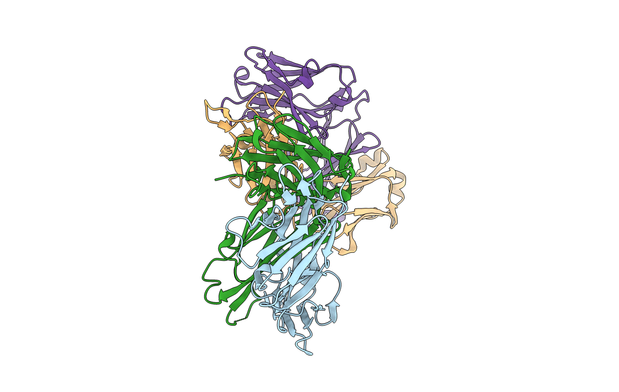
6KS2 image
Entry Detail
PDB ID:
6KS2
Keywords:
Title:
Structure of anti-Ghrelin receptor antibody
Biological Source:
Source Organism:
Host Organism:
PDB Version:
Deposition Date:
2019-08-23
Release Date:
2020-08-12
Method Details:
Experimental Method:
Resolution:
1.75 Å
R-Value Free:
0.23
R-Value Work:
0.19
R-Value Observed:
0.19
Space Group:
P 21 21 21
Macromolecular Entities
Polymer Type:polypeptide(L)
Description:Fab7881 Light Chain (FabL)
Chain IDs:A, C
Chain Length:219
Number of Molecules:2
Biological Source:Mus musculus
Polymer Type:polypeptide(L)
Description:Fab7881 Heavy Chain (FabH)
Chain IDs:B, D
Chain Length:218
Number of Molecules:2
Biological Source:Mus musculus
Primary Citation
Structure of an antagonist-bound ghrelin receptor reveals possible ghrelin recognition mode.
Nat Commun 11 4160 4160 (2020)
PMID: 32814772 DOI: 10.1038/s41467-020-17554-1

Abstact

Ghrelin is a gastric peptide hormone with important physiological functions. The unique feature of ghrelin is its Serine 3 acyl-modification, which is essential for ghrelin's activity. However, it remains to be elucidated why the acyl-modification of ghrelin is necessary for activity. To address these questions, we solved the crystal structure of the ghrelin receptor bound to antagonist. The ligand-binding pocket of the ghrelin receptor is bifurcated by a salt bridge between E124 and R283. A striking feature of the ligand-binding pocket of the ghrelin receptor is a wide gap (crevasse) between the TM6 and TM7 bundles that is rich in hydrophobic amino acids, including a cluster of phenylalanine residues. Mutagenesis analyses suggest that the interaction between the gap structure and the acyl acid moiety of ghrelin may participate in transforming the ghrelin receptor into an active conformation.

Legend

Protein

Chemical

Disease

Primary Citation of related structures