6KOB image
Entry Detail
PDB ID:
6KOB
Keywords:
Title:
X-ray Structure of the proton-pumping cytochrome aa3-600 menaquinol oxidase from Bacillus subtilis
Biological Source:
PDB Version:
Deposition Date:
2019-08-09
Release Date:
2020-01-15
Method Details:
Experimental Method:
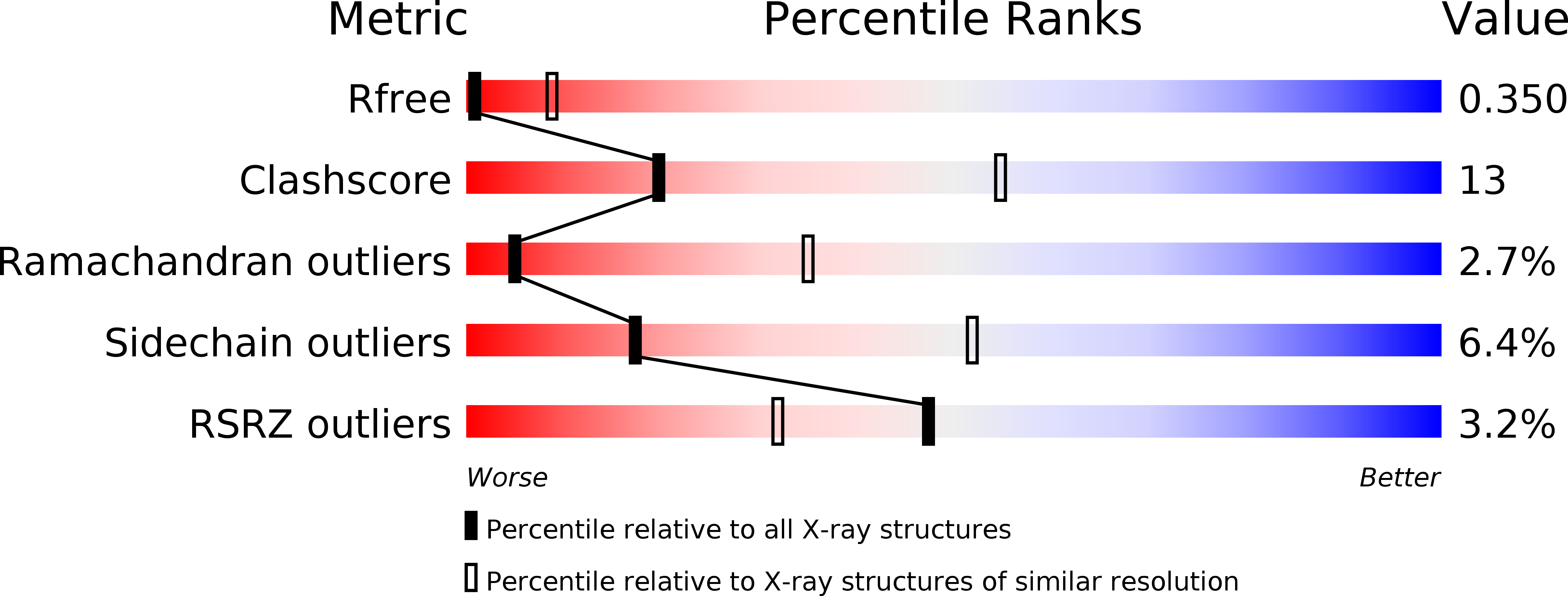
Resolution:
3.60 Å
R-Value Free:
0.35
R-Value Work:
0.32
Space Group:
P 1 21 1
Macromolecular Entities
Polymer Type:polypeptide(L)
Description:AA3-600 quinol oxidase subunit I
Chain IDs:A, H (auth: E)
Chain Length:655
Number of Molecules:2
Biological Source:Bacillus subtilis
Polymer Type:polypeptide(L)
Description:Quinol oxidase subunit 2
Chain IDs:B, G (auth: F)
Chain Length:296
Number of Molecules:2
Biological Source:Bacillus subtilis
Polymer Type:polypeptide(L)
Description:AA3-600 quinol oxidase subunit IIII
Chain IDs:C, E (auth: G)
Chain Length:204
Number of Molecules:2
Biological Source:Bacillus subtilis
Polymer Type:polypeptide(L)
Description:AA3-600 quinol oxidase subunit IV,Quinol oxidase subunit 4
Chain IDs:D, F (auth: H)
Chain Length:124
Number of Molecules:2
Biological Source:Bacillus subtilis (strain 168)
Primary Citation
Structure of the cytochromeaa3-600 heme-copper menaquinol oxidase bound to inhibitor HQNO shows TM0 is part of the quinol binding site.
Proc.Natl.Acad.Sci.USA 117 872 876 (2020)
PMID: 31888984 DOI: 10.1073/pnas.1915013117

Abstact

Virtually all proton-pumping terminal respiratory oxygen reductases are members of the heme-copper oxidoreductase superfamily. Most of these enzymes use reduced cytochrome c as a source of electrons, but a group of enzymes have evolved to directly oxidize membrane-bound quinols, usually menaquinol or ubiquinol. All of the quinol oxidases have an additional transmembrane helix (TM0) in subunit I that is not present in the related cytochrome c oxidases. The current work reports the 3.6-Å-resolution X-ray structure of the cytochrome aa3 -600 menaquinol oxidase from Bacillus subtilis containing 1 equivalent of menaquinone. The structure shows that TM0 forms part of a cleft to accommodate the menaquinol-7 substrate. Crystals which have been soaked with the quinol-analog inhibitor HQNO (N-oxo-2-heptyl-4-hydroxyquinoline) or 3-iodo-HQNO reveal a single binding site where the inhibitor forms hydrogen bonds to amino acid residues shown previously by spectroscopic methods to interact with the semiquinone state of menaquinone, a catalytic intermediate.

Legend

Protein

Chemical

Disease

Primary Citation of related structures