Deposition Date
2019-07-31
Release Date
2020-11-18
Last Version Date
2023-11-22
Entry Detail
PDB ID:
6KML
Keywords:
Title:
2.09 Angstrom resolution crystal structure of tetrameric HigBA toxin-antitoxin complex from E.coli
Biological Source:
Source Organism(s):
Escherichia coli K-12 (Taxon ID: 83333)
Expression System(s):
Method Details:
Experimental Method:
Resolution:
2.10 Å
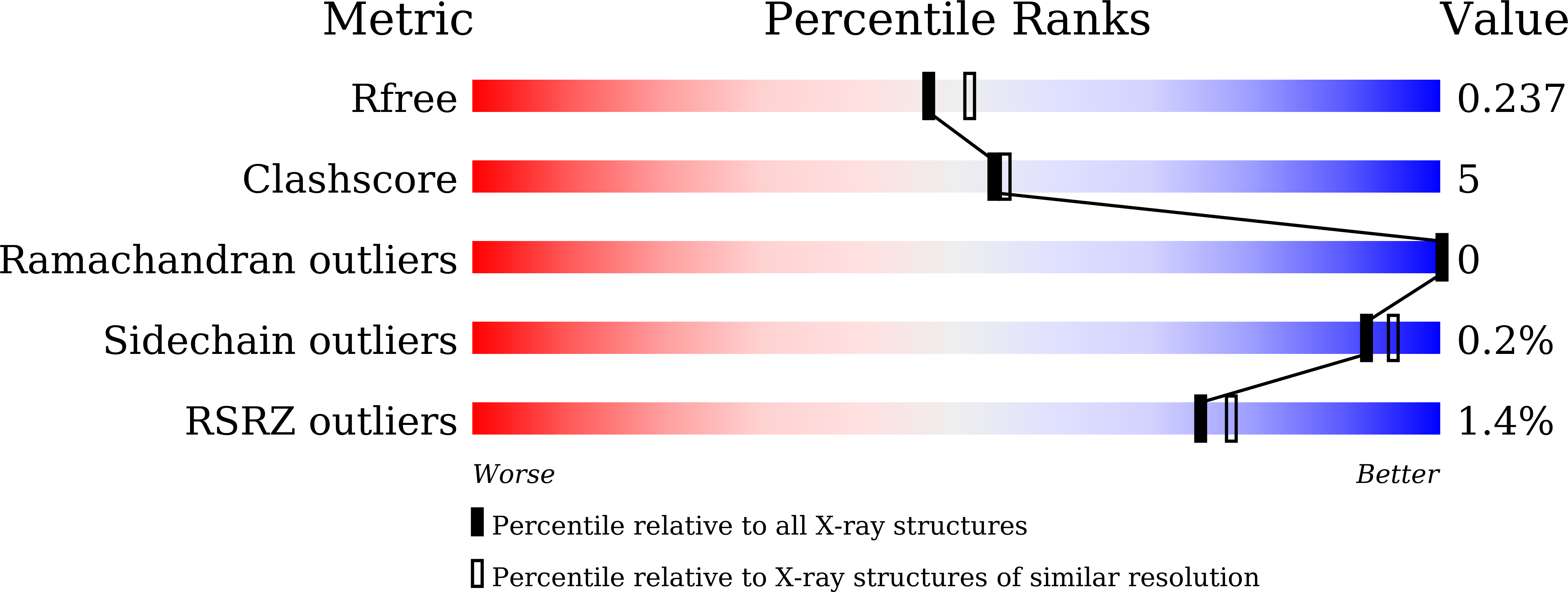
R-Value Free:
0.23
R-Value Work:
0.19
R-Value Observed:
0.19
Space Group:
P 1 21 1