6KLK image
Deposition Date 2019-07-30
Release Date 2019-11-06
Last Version Date 2023-11-22
Entry Detail
PDB ID:
6KLK
Keywords:
Title:
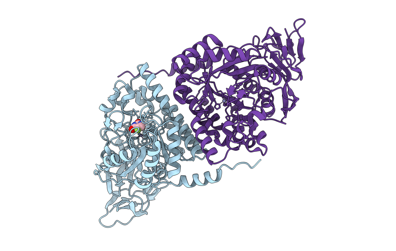
Crystal structure of the Pseudomonas aeruginosa dihydropyrimidinase complexed with 5-FU
Biological Source:
Method Details:
Experimental Method:
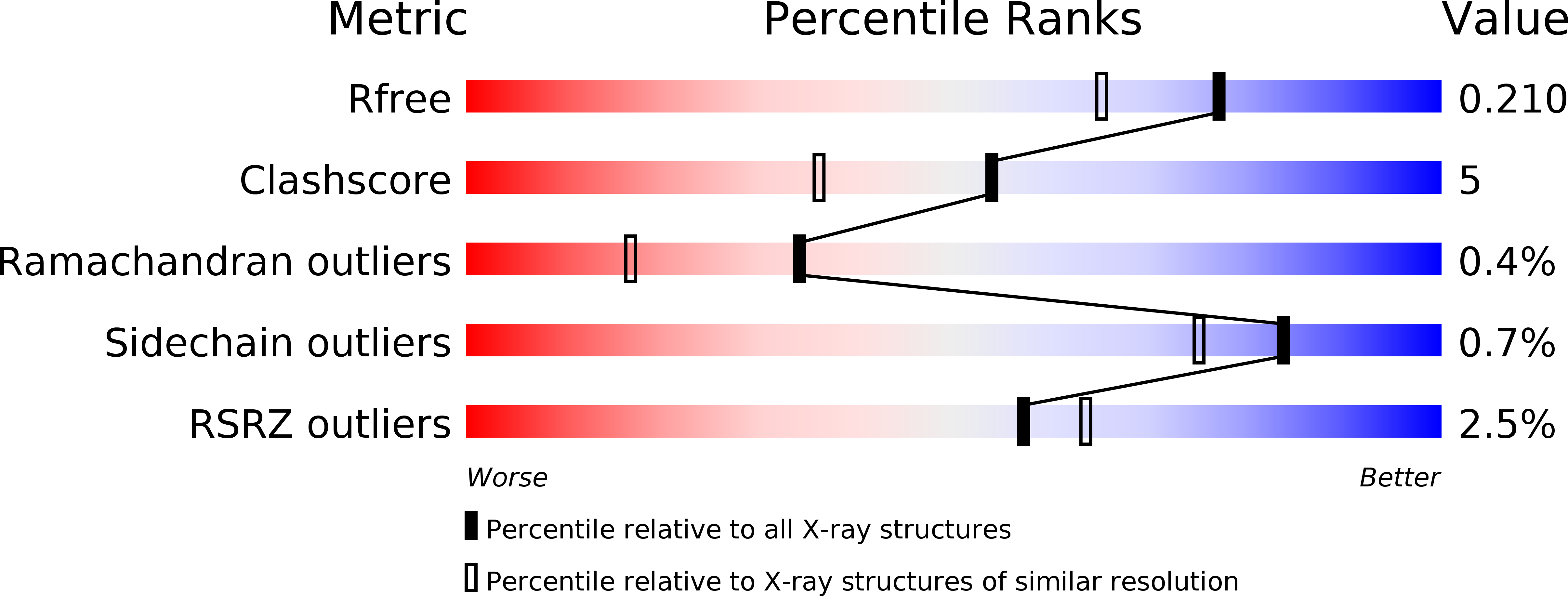
Resolution:
1.76 Å
R-Value Free:
0.21
R-Value Work:
0.18
R-Value Observed:
0.18
Space Group:
P 31 2 1
Macromolecular Entities
Structures with similar UniProt ID
Protein Blast
Polymer Type:polypeptide(L)
Molecule:D-hydantoinase/dihydropyrimidinase
Gene (Uniprot):dht
Chain IDs:A, B
Chain Length:479
Number of Molecules:2
Biological Source:Pseudomonas aeruginosa (strain ATCC 15692 / DSM 22644 / CIP 104116 / JCM 14847 / LMG 12228 / 1C / PRS 101 / PAO1)
Modified Residue
Compound ID Chain ID Parent Comp ID Details 2D Image
KCX A LYS modified residue
Primary Citation
Crystal structure of dihydropyrimidinase in complex with anticancer drug 5-fluorouracil.
Biochem.Biophys.Res.Commun. 519 160 165 (2019)
PMID: 31481233 DOI: 10.1016/j.bbrc.2019.08.153

Abstact

Dihydropyrimidinase (DHPase) catalyzes the reversible cyclization of dihydrouracil to N-carbamoyl-β-alanine in the second step of the pyrimidine degradation pathway. Whether 5-fluorouracil (5-FU), the best-known fluoropyrimidine that is used to target the enzyme thymidylate synthase for anticancer therapy, can bind to DHPase remains unknown. In this study, we found that 5-FU can form a stable complex with Pseudomonas aeruginosa DHPase (PaDHPase). The crystal structure of PaDHPase complexed with 5-FU was determined at 1.76 Å resolution (PDB entry 6KLK). Various interactions between 5-FU and PaDHPase were examined. Six residues, namely, His61, Tyr155, Asp316, Cys318, Ser289 and Asn337, of PaDHPase were involved in 5-FU binding. Except for Cys318, these residues are also known as the substrate-binding sites of DHPase. 5-FU interacts with the main chains of residues Ser289 (3.0 Å) and Asn337 (3.2 Å) and the side chains of residues Tyr155 (2.8 Å) and Cys318 (2.9 Å). Mutation at either Tyr155 or Cys318 of PaDHPase caused a low 5-FU binding activity of PaDHPase. This structure and the binding mode provided molecular insights into how the dimetal center in DHPase undergoes a conformational change during 5-FU binding. Further research can directly focus on revisiting the role of DHPase in anticancer therapy.

Legend

Protein

Chemical

Disease

Primary Citation of related structures
Feedback Form
Name
Email
Institute
Feedback