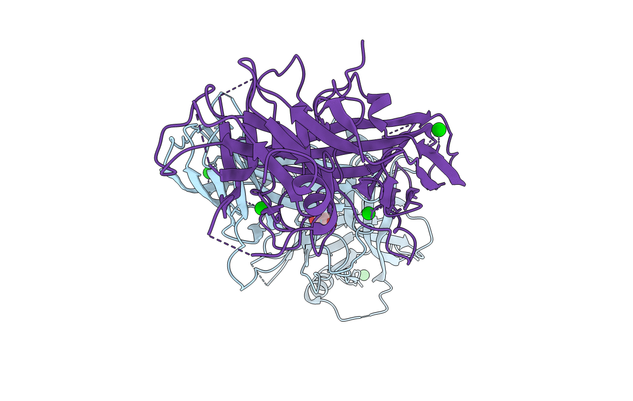
6KL7 image
Entry Detail
PDB ID:
6KL7
Title:
Beta-arrestin 1 mutant S13D/T275D
Biological Source:
Source Organism:
Host Organism:
PDB Version:
Deposition Date:
2019-07-29
Release Date:
2020-01-29
Method Details:
Experimental Method:
Resolution:
2.79 Å
R-Value Free:
0.27
R-Value Work:
0.21
R-Value Observed:
0.22
Space Group:
P 1 21 1
Macromolecular Entities
Polymer Type:polypeptide(L)
Description:Beta-arrestin-1
Mutations:S13D, T275D
Chain IDs:A, B
Chain Length:422
Number of Molecules:2
Biological Source:Rattus norvegicus
Primary Citation
Conformational Dynamics and Functional Implications of Phosphorylated beta-Arrestins.
Structure 28 314 ? (2020)
PMID: 31948726 DOI: 10.1016/j.str.2019.12.008

Abstact

Arrestins desensitize and/or internalize G-protein-coupled receptors by interacting with phosphorylated receptors. A few studies have reported that arrestins themselves can be phosphorylated, and the phosphorylation status modulates their cellular functions. However, the effects of phosphorylation on arrestin structure have not been studied. Here, we investigated the conformational changes in β-arrestin-1 and -2 upon incorporation of phospho-mimetic mutations into the known phosphorylation sites (i.e., S412D for β-arrestin-1 and S14D, T276D, S14D/T276D, S361D, T383D, and S361D/T383D for β-arrestin-2) by using hydrogen/deuterium-exchange mass spectrometry (HDX-MS). HDX-MS analysis suggested that β-arrestin-2 S14D/T276D shows an HDX profile similar to the pre-active states, resulting in increased interaction with receptors. Phospho-mimetic mutation at corresponding residues of β-arrestin-1 (i.e., S13D/T275D) induced similar conformational and functional consequences, and the detailed structural changes related to β-arrestin-1 S13D/T275D were investigated further by X-ray crystallography.

Legend

Protein

Chemical

Disease

Primary Citation of related structures