Deposition Date
2019-07-24
Release Date
2020-07-29
Last Version Date
2023-11-22
Entry Detail
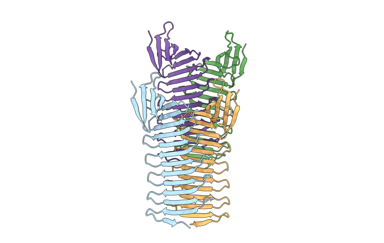
Biological Source:
Source Organism:
Borreliella burgdorferi (Taxon ID: 139)
Host Organism:
Method Details:
Experimental Method:
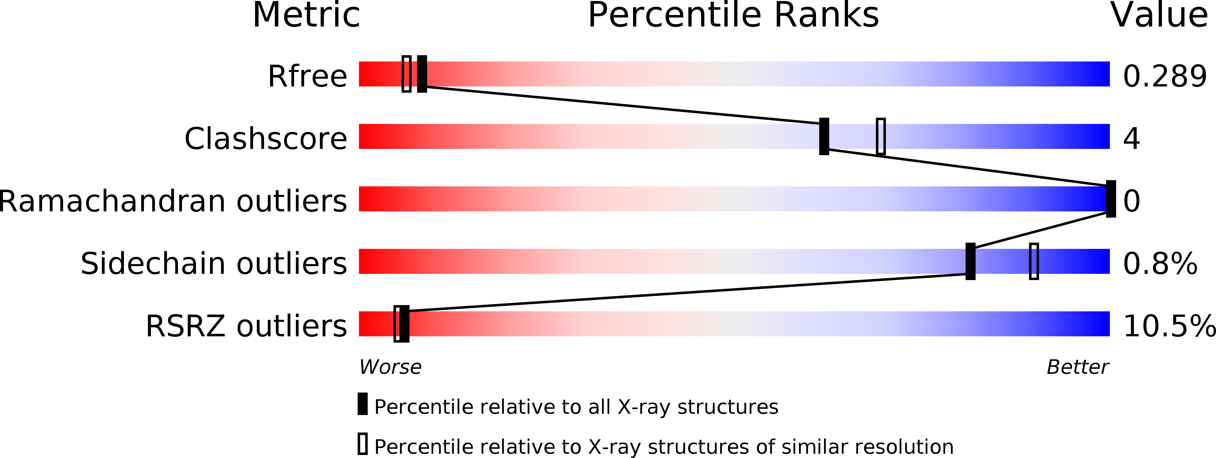
Resolution:
2.20 Å
R-Value Free:
0.28
R-Value Work:
0.22
R-Value Observed:
0.22
Space Group:
C 1 2 1