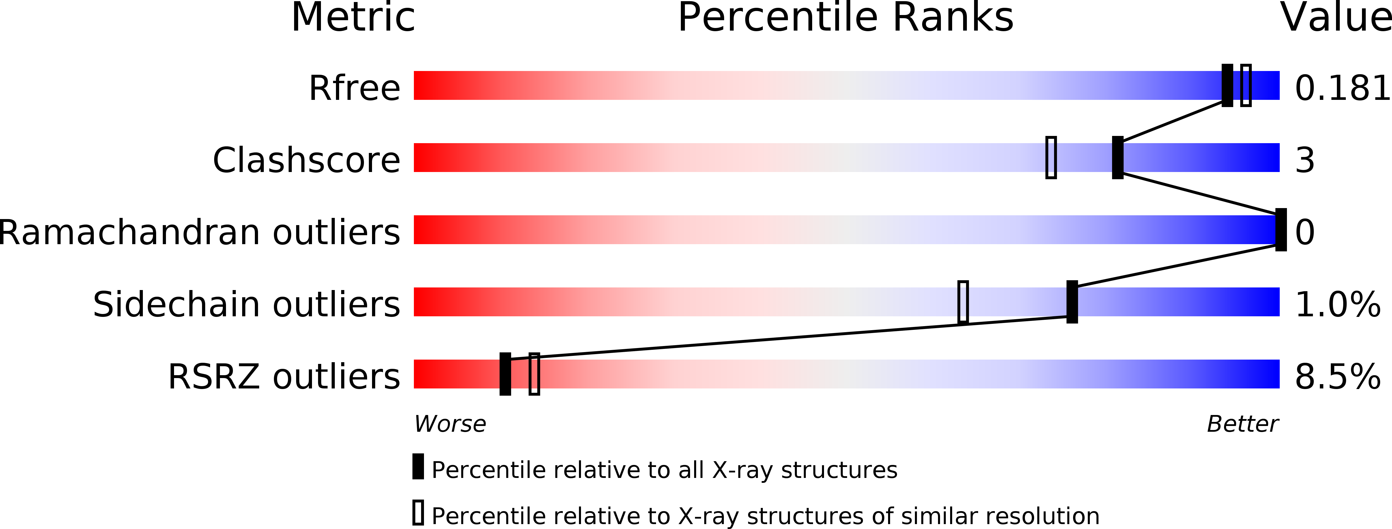
6KJU image
Deposition Date 2019-07-23
Release Date 2019-11-13
Last Version Date 2024-10-16
Entry Detail
PDB ID:
6KJU
Keywords:
Title:
Huge conformation shift of Vibrio cholerae VqmA dimer in the absence of target DNA provides insight into DNA-binding mechanisms of LuxR-type receptors
Biological Source:
Source Organism:
Vibrio cholerae (Taxon ID: 666)
Host Organism:
Method Details:
Experimental Method:
Resolution:
1.75 Å
R-Value Free:
0.19
R-Value Work:
0.17
R-Value Observed:
0.17
Space Group:
P 21 21 21
Macromolecular Entities
Structures with similar UniProt ID
Protein Blast
Polymer Type:polypeptide(L)
Molecule:Helix-turn-helix transcriptional regulator
Chain IDs:A, B
Chain Length:254
Number of Molecules:2
Biological Source:Vibrio cholerae
Ligand Molecules
Primary Citation
Large conformation shifts of Vibrio cholerae VqmA dimer in the absence of target DNA provide insight into DNA-binding mechanisms of LuxR-type receptors.
Biochem.Biophys.Res.Commun. 520 399 405 (2019)
PMID: 31606206 DOI: 10.1016/j.bbrc.2019.10.063

Abstact

Quorum sensing regulates the biofilm formation and expression of virulence factors in Vibrio cholerae, an obligate human pathogen that continues to imperil human health. Cytoplasmic transcription factor VqmA is a LuxR-type receptor ubiquitous in the Vibrio genus and one vibriophage VP882 and plays an important role in V. cholerae pathogenicity. Here we presented the X-ray crystal structure of V. cholerae VqmA-DPO complex and compared it with the previously determined VqmA-DPO-DNA complex. To our knowledge, this is the first report on the crystal structures of the same LuxR-type receptor with two conformations of binding to DNA and not binding to DNA. Based on the results of structural analysis and biochemical assays, we revealed the secondary structure of the linker region between two function domains changed significantly, and DNA binding domains were covalently linked by a disulfide bond formed by the highly conserved Cys134. Besides, the distance between two DBD monomers became longer than that in DNA-binding conformation, and two α8 helixes underwent a large conformation shift. The results of the structure-function analyses presented here improve our understanding of the complex mechanisms in the conformational changes of LuxR-type receptors caused by DNA binding.

Legend

Protein

Chemical

Disease

Primary Citation of related structures