6KHR image
Deposition Date 2019-07-16
Release Date 2019-12-18
Last Version Date 2023-11-22
Entry Detail
PDB ID:
6KHR
Keywords:
Title:
Structure of glycinamide-RNase-transformylase T from Mycobacterium tuberculosis
Biological Source:
Source Organism:
Host Organism:
Method Details:
Experimental Method:
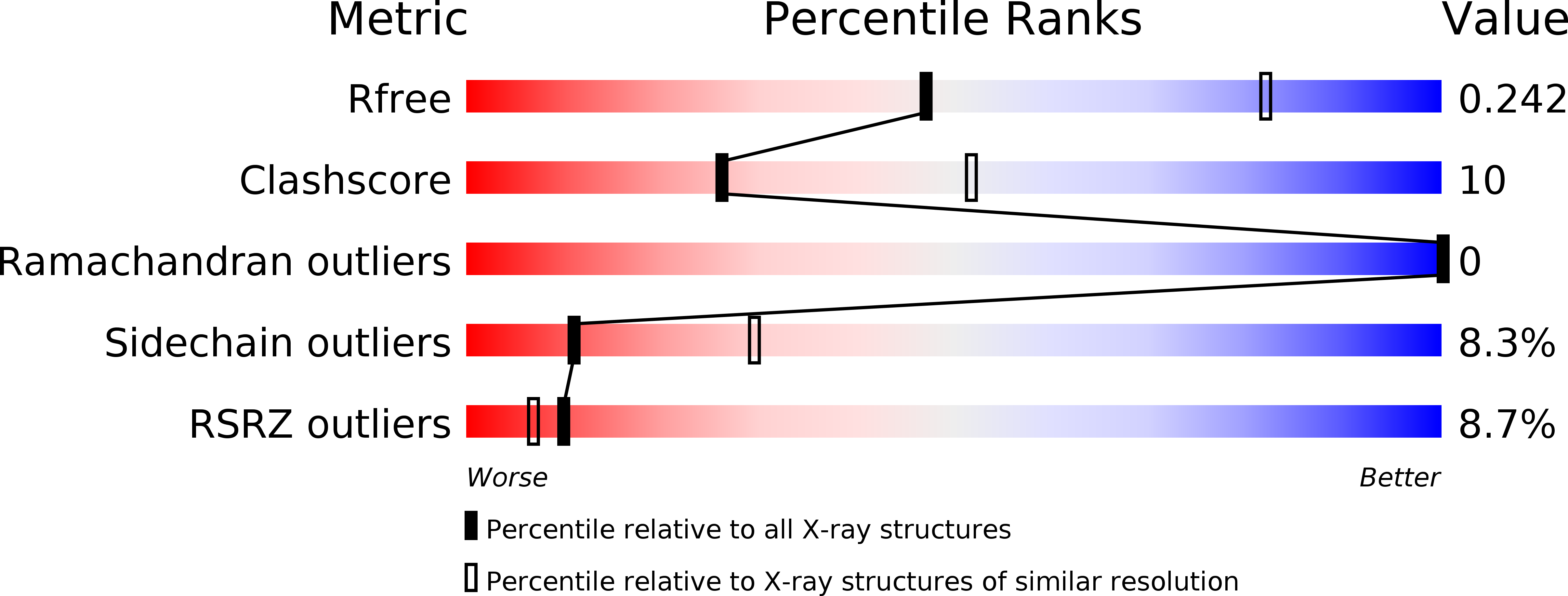
Resolution:
2.79 Å
R-Value Free:
0.24
R-Value Work:
0.21
R-Value Observed:
0.21
Space Group:
P 31 2 1
Macromolecular Entities
Structures with similar UniProt ID
Protein Blast
Polymer Type:polypeptide(L)
Molecule:Formate-dependent phosphoribosylglycinamide formyltransferase
Gene (Uniprot):purT
Chain IDs:A, B
Chain Length:420
Number of Molecules:2
Biological Source:Mycobacterium tuberculosis H37Rv
Primary Citation
Structural characterization of glycinamide-RNase-transformylase T fromMycobacterium tuberculosis.
Emerg Microbes Infect 9 58 66 (2020)
PMID: 31894729 DOI: 10.1080/22221751.2019.1707716

Abstact

Enzymes from the purine salvage pathway in Mycobacterium tuberculosis (Mtb) have been regarded as an attractive target for the development of anti-bacterial drugs. Although this pathway has not been extensively studied in Mtb, it has been identified as essential for growth and survival. Glycinamide-RNase-transformylase T (PurT) is found only in some specific bacteria including Mtb and utilizes ATP-dependent ligation to catalyze the formylation of 5'-phosphoribosyl-glycinamide (GAR) in the third reaction of the de novo purine salvage pathway. In the study, we determined the crystal structure of MtbPurT at a resolution of 2.79 Å. In contrast to Pyrococcus horikoshii OT3 PurT (phBCCPPurT), MtbPurT exhibits an "open" conformation, which results in a broader ATP-binding pocket and thus might facilitate the entry and exit of the cofactor. Additionally, active site superposition with E.coli PurT (EcPurT) showed that residues involved in the ATP-binding site in MtbPurT exhibited structural similarity but had notable difference in the GAR-binding site. The loop 383-389 in MtbPurT was much shorter and shifted 5.7 Å away from the phosphate of the GAR substrate. The different GAR-binding mode might result in a large conformational change in MtbPurT, and would provide a possible opportunity for anti-TB drug development.

Legend

Protein

Chemical

Disease

Primary Citation of related structures