6K60 image
Deposition Date 2019-05-31
Release Date 2019-11-27
Last Version Date 2024-11-13
Entry Detail
PDB ID:
6K60
Keywords:
Title:
Structural and functional basis for HLA-G isoform recognition of immune checkpoint receptor LILRBs
Biological Source:
Source Organism:
Homo sapiens (Taxon ID: 9606)
Host Organism:
Method Details:
Experimental Method:
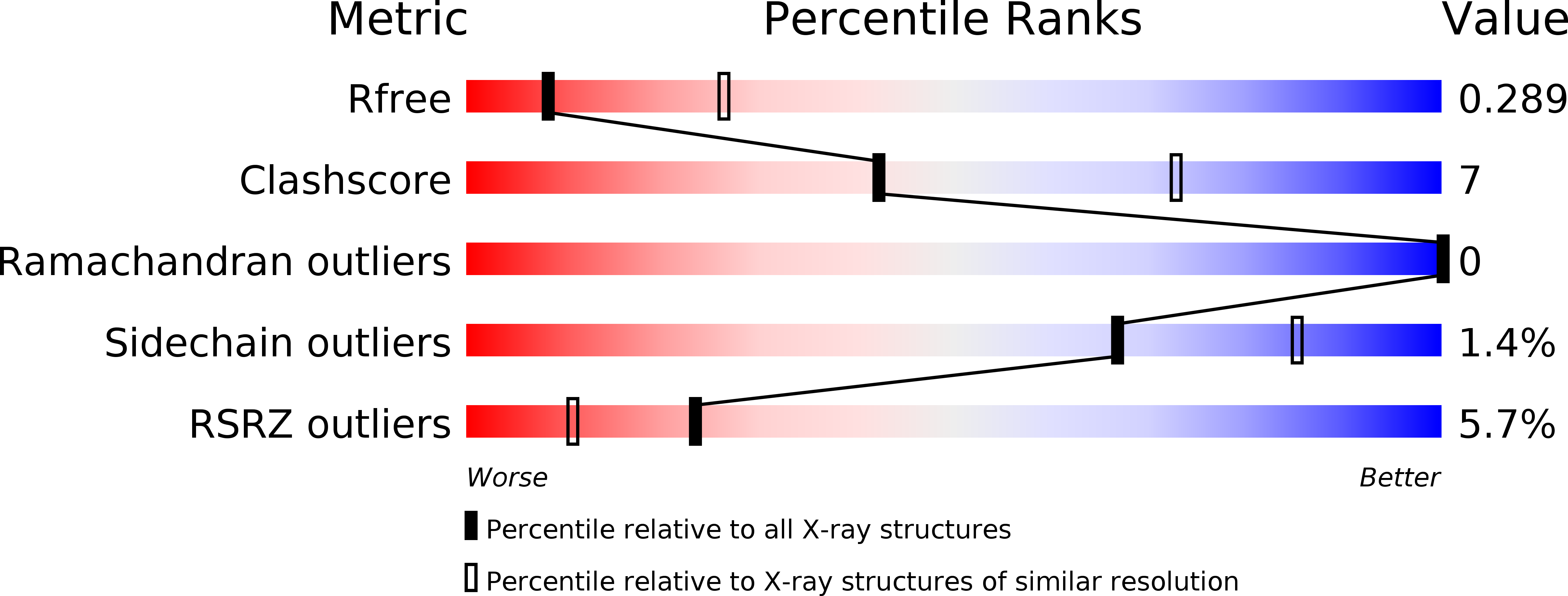
Resolution:
3.15 Å
R-Value Free:
0.28
R-Value Work:
0.26
R-Value Observed:
0.26
Space Group:
H 3 2
Macromolecular Entities
Structures with similar UniProt ID
Protein Blast
Polymer Type:polypeptide(L)
Molecule:HLA class I histocompatibility antigen, alpha chain G
Gene (Uniprot):HLA-G
Mutagens:C42S
Chain IDs:A, E
Chain Length:277
Number of Molecules:2
Biological Source:Homo sapiens
Structures with similar UniProt ID
Protein Blast
Polymer Type:polypeptide(L)
Molecule:Beta-2-microglobulin
Gene (Uniprot):B2M
Chain IDs:B, F
Chain Length:100
Number of Molecules:2
Biological Source:Homo sapiens
Structures with similar UniProt ID
Protein Blast
Polymer Type:polypeptide(L)
Molecule:Peptide from Histone H2A.J
Chain IDs:C, G
Chain Length:9
Number of Molecules:2
Biological Source:Homo sapiens
Structures with similar UniProt ID
Protein Blast
Polymer Type:polypeptide(L)
Molecule:Leukocyte immunoglobulin-like receptor subfamily B member 1
Chain IDs:D, H
Chain Length:198
Number of Molecules:2
Biological Source:Homo sapiens
Ligand Molecules
Primary Citation
Structural and Functional Basis for LILRB Immune Checkpoint Receptor Recognition of HLA-G Isoforms.
J Immunol. 203 3386 3394 (2019)
PMID: 31694909 DOI: 10.4049/jimmunol.1900562

Abstact

Human leukocyte Ig-like receptors (LILR) LILRB1 and LILRB2 are immune checkpoint receptors that regulate a wide range of physiological responses by binding to diverse ligands, including HLA-G. HLA-G is exclusively expressed in the placenta, some immunoregulatory cells, and tumors and has several unique isoforms. However, the recognition of HLA-G isoforms by LILRs is poorly understood. In this study, we characterized LILR binding to the β2-microglobulin (β2m)-free HLA-G1 isoform, which is synthesized by placental trophoblast cells and tends to dimerize and multimerize. The multimerized β2m-free HLA-G1 dimer lacked detectable affinity for LILRB1, but bound strongly to LILRB2. We also determined the crystal structure of the LILRB1 and HLA-G1 complex, which adopted the typical structure of a classical HLA class I complex. LILRB1 exhibits flexible binding modes with the α3 domain, but maintains tight contacts with β2m, thus accounting for β2m-dependent binding. Notably, both LILRB1 and B2 are oriented at suitable angles to permit efficient signaling upon complex formation with HLA-G1 dimers. These structural and functional features of ligand recognition by LILRs provide novel insights into their important roles in the biological regulations.

Legend

Protein

Chemical

Disease

Primary Citation of related structures
Feedback Form
Name
Email
Institute
Feedback